2WYQ | pdb_00002wyq

THE CRYSTAL STRUCTURE OF THE UBIQUITIN-LIKE (UBL) DOMAIN OF HHR23A (HUMAN HOMOLOGUE A OF RAD23)


Experimental Data Snapshot

  • Method: X-RAY DIFFRACTION
  • Resolution: 1.65 Å
  • R-Value Free: 
    0.160 (Depositor), 0.236 (DCC) 
  • R-Value Work: 
    0.124 (Depositor) 
  • R-Value Observed: 
    0.126 (Depositor) 

Starting Model: experimental
View more details

wwPDB Validation 3D Report Full Report

Validation slider image for 2WYQ

This is version 1.3 of the entry. See complete history

Literature

The Crystal Structure of the Ubiquitin-Like (Ubl) Domain of Human Homologue a of Rad23 (Hhr23A) Protein

Chen, Y.W.Tajima, T.Agrawal, S.

(2011) Protein Eng Des Sel 24: 131

  • DOI: https://doi.org/10.1093/protein/gzq084
  • Primary Citation Related Structures: 
    2WYQ

  • PubMed Abstract: 

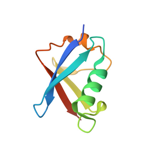
    The human homologue of the yeast Rad23 protein, hHR23A, plays dual roles in DNA repair as well as in translocating polyubiquitinated proteins to the proteasome. We determined the three-dimensional structure of its ubiquitin-like (UbL) domain by X-ray crystallography. It has the same overall structure and fold characteristics as ubiquitin and other members of the UbL domain family, with overall root mean square deviations in Cα positions in the range of 1.0-1.3 Å. There are local differences in the α1-β3 loop where hHR23A UbL domain has three more residues constituting a bigger loop. Analysis of the crystal packing revealed a possible dimeric arrangement mediated by the three residues (Leu10, Ile49 and Met75) that are known to be critical for molecular interactions. In contrast to the overall well-defined structure, these three residues are either disordered or have multiple conformations, suggesting that conformation variability is an important property of the binding surface. The electrostatic potentials at the binding surface are conserved among the family, with the hHR23B domain being the most similar to this structure. The intra-molecular complexes formed by the UbL domain of hHR23A with its UbA1 or UbA2 domains was studied by comparative homology modelling, which suggests these two interactions are structurally similar and are mutually exclusive.


  • Organizational Affiliation
    • King's College London, Randall Division of Cell and Molecular Biophysics, New Hunt's House, Guy's Campus, London SE11UL, UK. yu-wai.chen@kcl.ac.uk

Macromolecule Content 

  • Total Structure Weight: 9.77 kDa 
  • Atom Count: 771 
  • Modeled Residue Count: 77 
  • Deposited Residue Count: 85 
  • Unique protein chains: 1

Macromolecules

Find similar proteins by:|  3D Structure
Entity ID: 1
MoleculeChains  Sequence LengthOrganismDetailsImage
UV EXCISION REPAIR PROTEIN RAD23 HOMOLOG A85Homo sapiensMutation(s): 0 
EC: 1.14.11
UniProt & NIH Common Fund Data Resources
Find proteins for P54725 (Homo sapiens)
Explore P54725 
Go to UniProtKB:  P54725
PHAROS:  P54725
GTEx:  ENSG00000179262 
Entity Groups
Sequence Clusters30% Identity50% Identity70% Identity90% Identity95% Identity100% Identity
UniProt GroupP54725
Sequence Annotations
Expand
Reference Sequence

Experimental Data & Validation

Experimental Data

  • Method: X-RAY DIFFRACTION
  • Resolution: 1.65 Å
  • R-Value Free:  0.160 (Depositor), 0.236 (DCC) 
  • R-Value Work:  0.124 (Depositor) 
  • R-Value Observed: 0.126 (Depositor) 
Space Group: P 65 2 2
Unit Cell:
Length ( Å )Angle ( ˚ )
a = 78.434α = 90
b = 78.434β = 90
c = 63.472γ = 120
Software Package:
Software NamePurpose
REFMACrefinement
MOSFLMdata reduction
SCALAdata scaling
PHASERphasing

Structure Validation

View Full Validation Report


Entry History 

Deposition Data

  • Released Date: 2010-11-10 
  • Deposition Author(s): Chen, Y.W.

Revision History  (Full details and data files)

  • Version 1.0: 2010-11-10
    Type: Initial release
  • Version 1.1: 2011-05-08
    Changes: Version format compliance
  • Version 1.2: 2011-07-13
    Changes: Version format compliance
  • Version 1.3: 2023-12-20
    Changes: Data collection, Database references, Derived calculations, Other, Refinement description