2WYA | pdb_00002wya

CRYSTAL STRUCTURE OF HUMAN MITOCHONDRIAL 3-HYDROXY-3-METHYLGLUTARYL- COENZYME A SYNTHASE 2 (HMGCS2)


Experimental Data Snapshot

  • Method: X-RAY DIFFRACTION
  • Resolution: 1.70 Å
  • R-Value Free: 
    0.184 (Depositor), 0.177 (DCC) 
  • R-Value Work: 
    0.161 (Depositor), 0.155 (DCC) 
  • R-Value Observed: 
    0.162 (Depositor) 

Starting Model: experimental
View more details

wwPDB Validation 3D Report Full Report

Validation slider image for 2WYA

Ligand Structure Quality Assessment 


This is version 1.4 of the entry. See complete history

Literature

Crystal Structures of Human Hmg-Coa Synthase Isoforms Provide Insights Into Inherited Ketogenesis Disorders and Inhibitor Design.

Shafqat, N.Turnbull, A.Zschocke, J.Oppermann, U.Yue, W.W.

(2010) J Mol Biology 398: 497

  • DOI: https://doi.org/10.1016/j.jmb.2010.03.034
  • Primary Citation Related Structures: 
    2P8U, 2WYA

  • PubMed Abstract: 

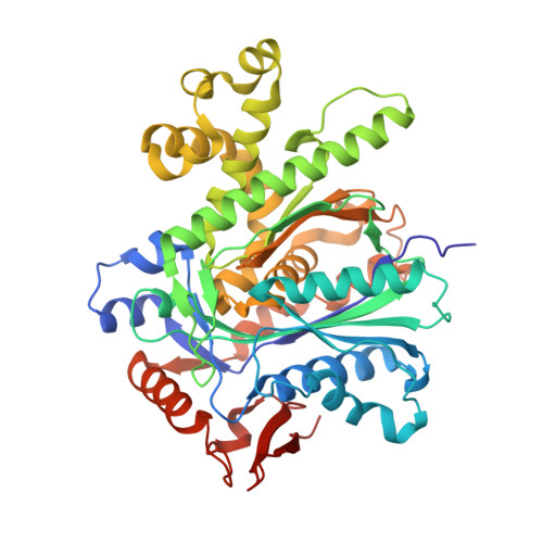
    3-Hydroxy-3-methylglutaryl coenzyme A (CoA) synthase (HMGCS) catalyzes the condensation of acetyl-CoA and acetoacetyl-CoA into 3-hydroxy-3-methylglutaryl CoA. It is ubiquitous across the phylogenetic tree and is broadly classified into three classes. The prokaryotic isoform is essential in Gram-positive bacteria for isoprenoid synthesis via the mevalonate pathway. The eukaryotic cytosolic isoform also participates in the mevalonate pathway but its end product is cholesterol. Mammals also contain a mitochondrial isoform; its deficiency results in an inherited disorder of ketone body formation. Here, we report high-resolution crystal structures of the human cytosolic (hHMGCS1) and mitochondrial (hHMGCS2) isoforms in binary product complexes. Our data represent the first structures solved for human HMGCS and the mitochondrial isoform, allowing for the first time structural comparison among the three isoforms. This serves as a starting point for the development of isoform-specific inhibitors that have potential cholesterol-reducing and antibiotic applications. In addition, missense mutations that cause mitochondrial HMGCS deficiency have been mapped onto the hHMGCS2 structure to rationalize the structural basis for the disease pathology.


  • Organizational Affiliation
    • Structural Genomics Consortium, University of Oxford, Oxford OX3 7DU, UK.

Macromolecule Content 

  • Total Structure Weight: 208.98 kDa 
  • Atom Count: 16,074 
  • Modeled Residue Count: 1,840 
  • Deposited Residue Count: 1,840 
  • Unique protein chains: 1

Macromolecules

Find similar proteins by:|  3D Structure
Entity ID: 1
MoleculeChains  Sequence LengthOrganismDetailsImage
HYDROXYMETHYLGLUTARYL-COA SYNTHASE, MITOCHONDRIAL
A, B, C, D
460Homo sapiensMutation(s): 0 
EC: 2.3.3.10
UniProt & NIH Common Fund Data Resources
Find proteins for P54868 (Homo sapiens)
Explore P54868 
Go to UniProtKB:  P54868
PHAROS:  P54868
GTEx:  ENSG00000134240 
Entity Groups
Sequence Clusters30% Identity50% Identity70% Identity90% Identity95% Identity100% Identity
UniProt GroupP54868
Sequence Annotations
Expand
Reference Sequence

Experimental Data & Validation

Experimental Data

  • Method: X-RAY DIFFRACTION
  • Resolution: 1.70 Å
  • R-Value Free:  0.184 (Depositor), 0.177 (DCC) 
  • R-Value Work:  0.161 (Depositor), 0.155 (DCC) 
  • R-Value Observed: 0.162 (Depositor) 
Space Group: P 1
Unit Cell:
Length ( Å )Angle ( ˚ )
a = 66.41α = 100
b = 83.514β = 108.08
c = 101.442γ = 96.3
Software Package:
Software NamePurpose
PHENIXrefinement
MOSFLMdata reduction
SCALAdata scaling
PHASERphasing

Structure Validation

View Full Validation Report


Ligand Structure Quality Assessment 


Entry History 

Deposition Data

Revision History  (Full details and data files)

  • Version 1.0: 2009-11-24
    Type: Initial release
  • Version 1.1: 2011-05-08
    Changes: Version format compliance
  • Version 1.2: 2011-07-13
    Changes: Version format compliance
  • Version 1.3: 2023-12-20
    Changes: Data collection, Database references, Derived calculations, Other, Refinement description
  • Version 1.4: 2024-11-20
    Changes: Structure summary