2WQV

Internalin domain of Listeria monocytogenes InlB: rhombohedral crystal form


Experimental Data Snapshot

  • Method: X-RAY DIFFRACTION
  • Resolution: 2.80 Å
  • R-Value Free: 0.238 
  • R-Value Work: 0.203 
  • R-Value Observed: 0.205 

wwPDB Validation   3D Report Full Report


This is version 1.3 of the entry. See complete history


Literature

Ligand-Mediated Dimerization of the met Receptor Tyrosine Kinase by the Bacterial Invasion Protein Inlb.

Ferraris, D.M.Gherardi, E.Di, Y.Heinz, D.W.Niemann, H.H.

(2010) J Mol Biol 395: 522

  • DOI: https://doi.org/10.1016/j.jmb.2009.10.074
  • Primary Citation of Related Structures:  
    2WQU, 2WQV, 2WQW, 2WQX

  • PubMed Abstract: 

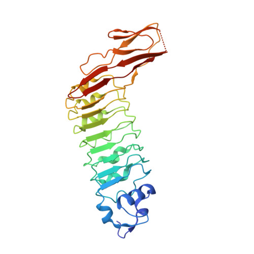
    The Listeria monocytogenes surface protein InlB mediates bacterial invasion into host cells by activating the human receptor tyrosine kinase Met. So far, it is unknown how InlB or the physiological Met ligand hepatocyte growth factor/scatter factor causes Met dimerization, which is considered a prerequisite for receptor activation. We determined two new structures of InlB, revealing a recurring, antiparallel, dimeric arrangement, in which the two protomers interact through the convex face of the leucine-rich repeat domain. The same contact is found in one structure of the InlB-Met complex. Mutations disrupting the interprotomeric contact of InlB reduced its ability to activate Met and downstream signaling. Conversely, stabilization of this crystal contact by two intermolecular disulfide bonds generates a constitutively dimeric InlB variant with exceptionally high signaling activity, which can stimulate cell motility and cell division. These data demonstrate that the signaling-competent InlB-Met complex assembles with 2:2 stoichiometry around a back-to-back InlB dimer, enabling the direct contact between the stalk region of two Met molecules.


  • Organizational Affiliation

    Division of Structural Biology, Helmholtz Centre for Infection Research (HZI), Inhoffenstrasse 7, 38124 Braunschweig, Germany. yyoshiki@riken.jp


Macromolecules
Find similar proteins by:  (by identity cutoff)  |  3D Structure
Entity ID: 1
MoleculeChains Sequence LengthOrganismDetailsImage
INTERNALIN B
A, B
289Listeria monocytogenes EGD-eMutation(s): 0 
UniProt
Find proteins for P0DQD2 (Listeria monocytogenes serovar 1/2a (strain ATCC BAA-679 / EGD-e))
Explore P0DQD2 
Go to UniProtKB:  P0DQD2
Entity Groups  
Sequence Clusters30% Identity50% Identity70% Identity90% Identity95% Identity100% Identity
UniProt GroupP0DQD2
Sequence Annotations
Expand
  • Reference Sequence
Experimental Data & Validation

Experimental Data

  • Method: X-RAY DIFFRACTION
  • Resolution: 2.80 Å
  • R-Value Free: 0.238 
  • R-Value Work: 0.203 
  • R-Value Observed: 0.205 
  • Space Group: H 3 2
Unit Cell:
Length ( Å )Angle ( ˚ )
a = 185.9α = 90
b = 185.9β = 90
c = 115.07γ = 120
Software Package:
Software NamePurpose
REFMACrefinement
XDSdata reduction
XSCALEdata scaling
PHASERphasing

Structure Validation

View Full Validation Report


Entry History 

Deposition Data

Revision History  (Full details and data files)

  • Version 1.0: 2009-11-10
    Type: Initial release
  • Version 1.1: 2011-07-13
    Changes: Advisory, Version format compliance
  • Version 1.2: 2019-03-06
    Changes: Data collection, Experimental preparation, Other
  • Version 1.3: 2023-12-20
    Changes: Data collection, Database references, Derived calculations, Other, Refinement description