2WQB | pdb_00002wqb

Structure of the Tie2 kinase domain in complex with a thiazolopyrimidine inhibitor


Experimental Data Snapshot

  • Method: X-RAY DIFFRACTION
  • Resolution: 2.95 Å
  • R-Value Free: 
    0.259 (Depositor), 0.250 (DCC) 
  • R-Value Work: 
    0.196 (Depositor), 0.193 (DCC) 
  • R-Value Observed: 
    0.199 (Depositor) 

Starting Model: experimental
View more details

wwPDB Validation 3D Report Full Report

Validation slider image for 2WQB

Ligand Structure Quality Assessment 


This is version 1.5 of the entry. See complete history

Literature

Novel Thienopyrimidine and Thiazolopyrimidine Kinase Inhibitors with Activity Against Tie-2 in Vitro and in Vivo.

Luke, R.W.Ballard, P.Buttar, D.Campbell, L.Curwen, J.Emery, S.C.Griffen, A.M.Hassall, L.Hayter, B.R.Jones, C.D.Mccoull, W.Mellor, M.Swain, M.L.Tucker, J.A.

(2009) Bioorg Med Chem Lett 19: 6670

  • DOI: https://doi.org/10.1016/j.bmcl.2009.10.001
  • Primary Citation Related Structures: 
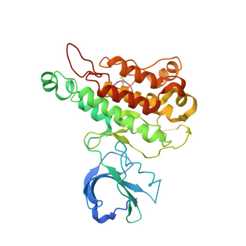
    2WQB

  • PubMed Abstract: 

    The SAR and improvement in potency against Tie2 of novel thienopyrimidine and thiazolopyrimidine kinase inhibitors are reported. The crystal structure of one of these compounds bound to the Tie-2 kinase domain is consistent with the SAR. These compounds have moderate potency in cellular assays of Tie-2 inhibition, good physical properties, DMPK, and show evidence of in vivo inhibition of Tie-2.


  • Organizational Affiliation
    • Cancer and Infection Research Area, AstraZeneca, Alderley Park, Macclesfield SK10 4TG, UK. richard.luke@astrazeneca.com

Macromolecule Content 

  • Total Structure Weight: 37.38 kDa 
  • Atom Count: 2,333 
  • Modeled Residue Count: 289 
  • Deposited Residue Count: 324 
  • Unique protein chains: 1

Macromolecules

Find similar proteins by:|  3D Structure
Entity ID: 1
MoleculeChains  Sequence LengthOrganismDetailsImage
ANGIOPOIETIN-1 RECEPTOR324Homo sapiensMutation(s): 1 
EC: 2.7.10.1
UniProt & NIH Common Fund Data Resources
Find proteins for Q02763 (Homo sapiens)
Explore Q02763 
Go to UniProtKB:  Q02763
PHAROS:  Q02763
GTEx:  ENSG00000120156 
Entity Groups
Sequence Clusters30% Identity50% Identity70% Identity90% Identity95% Identity100% Identity
UniProt GroupQ02763
Sequence Annotations
Expand
Reference Sequence

Small Molecules

Ligands 1 Unique
IDChains Name / Formula / InChI Key2D Diagram3D Interactions
QQ1

Query on QQ1


Download:Ideal Coordinates CCD File
B [auth A]2-[3-(CYCLOHEXYLMETHYL)-5-PHENYL-IMIDAZOL-4-YL]-[1,3]THIAZOLO[4,5-E]PYRIMIDIN-7-AMINE
C21 H22 N6 S
FACMGTJQTMTYEF-UHFFFAOYSA-N

Experimental Data & Validation

Experimental Data

  • Method: X-RAY DIFFRACTION
  • Resolution: 2.95 Å
  • R-Value Free:  0.259 (Depositor), 0.250 (DCC) 
  • R-Value Work:  0.196 (Depositor), 0.193 (DCC) 
  • R-Value Observed: 0.199 (Depositor) 
Space Group: C 2 2 21
Unit Cell:
Length ( Å )Angle ( ˚ )
a = 80.164α = 90
b = 108.791β = 90
c = 101.573γ = 90
Software Package:
Software NamePurpose
REFMACrefinement
MOSFLMdata reduction
SCALAdata scaling
AMoREphasing

Structure Validation

View Full Validation Report


Ligand Structure Quality Assessment 


Entry History 

Revision History  (Full details and data files)

  • Version 1.0: 2009-11-03
    Type: Initial release
  • Version 1.1: 2011-07-13
    Changes: Advisory, Refinement description, Version format compliance
  • Version 1.2: 2013-04-17
    Changes: Derived calculations, Refinement description
  • Version 1.3: 2019-03-06
    Changes: Data collection, Experimental preparation, Other
  • Version 1.4: 2023-12-20
    Changes: Data collection, Database references, Derived calculations, Other, Refinement description
  • Version 1.5: 2024-11-13
    Changes: Structure summary