2WQ9 | pdb_00002wq9

Crystal Structure of RBP4 bound to Oleic Acid


Experimental Data Snapshot

  • Method: X-RAY DIFFRACTION
  • Resolution: 1.65 Å
  • R-Value Free: 
    0.219 (Depositor), 0.218 (DCC) 
  • R-Value Work: 
    0.188 (Depositor), 0.186 (DCC) 
  • R-Value Observed: 
    0.189 (Depositor) 

Starting Model: experimental
View more details

wwPDB Validation 3D Report Full Report

Validation slider image for 2WQ9

Ligand Structure Quality Assessment 


This is version 1.6 of the entry. See complete history

Literature

Crystal Structure of Rbp4 Bound to Oleic Acid

Nanao, M.Stout, T.J.

To be published.

Macromolecule Content 

  • Total Structure Weight: 20.79 kDa 
  • Atom Count: 1,707 
  • Modeled Residue Count: 173 
  • Deposited Residue Count: 174 
  • Unique protein chains: 1

Macromolecules

Find similar proteins by:|  3D Structure
Entity ID: 1
MoleculeChains  Sequence LengthOrganismDetailsImage
RETINOL-BINDING PROTEIN 4174Homo sapiensMutation(s): 0 
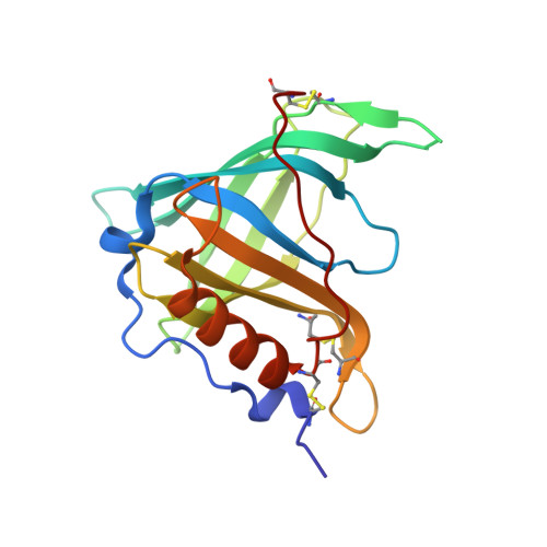
UniProt & NIH Common Fund Data Resources
Find proteins for P02753 (Homo sapiens)
Explore P02753 
Go to UniProtKB:  P02753
PHAROS:  P02753
GTEx:  ENSG00000138207 
Entity Groups
Sequence Clusters30% Identity50% Identity70% Identity90% Identity95% Identity100% Identity
UniProt GroupP02753
Sequence Annotations
Expand
Reference Sequence

Small Molecules

Ligands 3 Unique
IDChains Name / Formula / InChI Key2D Diagram3D Interactions
OLA

Query on OLA


Download:Ideal Coordinates CCD File
F [auth A]OLEIC ACID
C18 H34 O2
ZQPPMHVWECSIRJ-KTKRTIGZSA-N
GOL

Query on GOL


Download:Ideal Coordinates CCD File
G [auth A],
H [auth A],
I [auth A]
GLYCEROL
C3 H8 O3
PEDCQBHIVMGVHV-UHFFFAOYSA-N
CL

Query on CL


Download:Ideal Coordinates CCD File
B [auth A],
C [auth A],
D [auth A],
E [auth A],
J [auth A]
CHLORIDE ION
Cl
VEXZGXHMUGYJMC-UHFFFAOYSA-M

Experimental Data & Validation

Experimental Data

  • Method: X-RAY DIFFRACTION
  • Resolution: 1.65 Å
  • R-Value Free:  0.219 (Depositor), 0.218 (DCC) 
  • R-Value Work:  0.188 (Depositor), 0.186 (DCC) 
  • R-Value Observed: 0.189 (Depositor) 
Space Group: H 3
Unit Cell:
Length ( Å )Angle ( ˚ )
a = 103.06α = 90
b = 103.06β = 90
c = 72.71γ = 120
Software Package:
Software NamePurpose
REFMACrefinement
XDSdata reduction
XSCALEdata scaling
PHASERphasing

Structure Validation

View Full Validation Report


Ligand Structure Quality Assessment 


Entry History 

Deposition Data

Revision History  (Full details and data files)

  • Version 1.0: 2010-09-01
    Type: Initial release
  • Version 1.1: 2011-05-08
    Changes: Version format compliance
  • Version 1.2: 2011-07-13
    Changes: Version format compliance
  • Version 1.3: 2017-06-28
    Changes: Data collection
  • Version 1.4: 2019-04-03
    Changes: Data collection, Source and taxonomy
  • Version 1.5: 2023-12-20
    Changes: Data collection, Database references, Derived calculations, Other, Refinement description
  • Version 1.6: 2024-10-23
    Changes: Structure summary