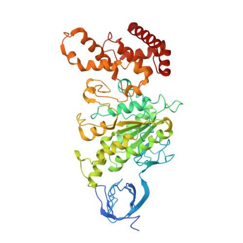
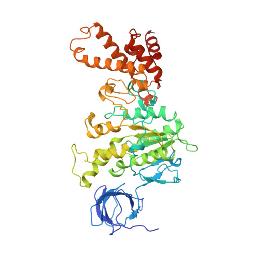
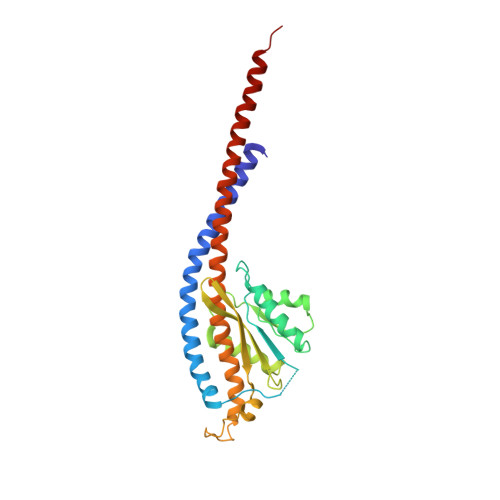
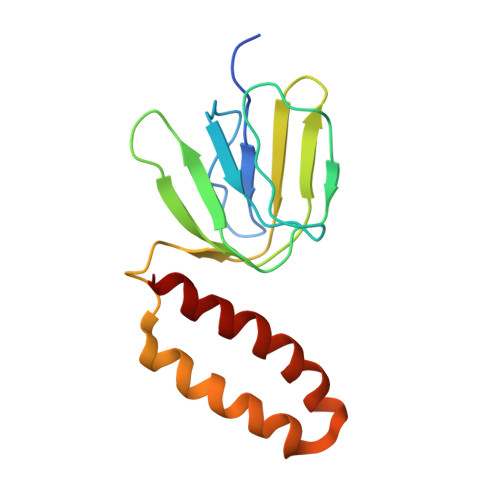
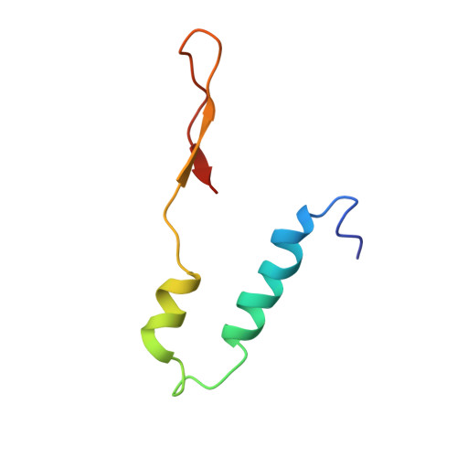
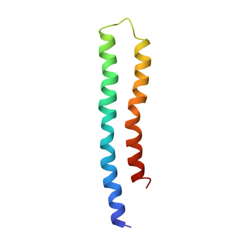
2WPD

The Mg.ADP inhibited state of the yeast F1c10 ATP synthase


Experimental Data Snapshot

  • Method: X-RAY DIFFRACTION
  • Resolution: 3.43 Å
  • R-Value Free: 0.297 
  • R-Value Work: 0.286 
  • R-Value Observed: 0.287 

Starting Models: experimental
View more details

wwPDB Validation   3D Report Full Report


Ligand Structure Quality Assessment 


This is version 1.3 of the entry. See complete history


Literature

Crystal Structure of the Mg.Adp-Inhibited State of the Yeast F1C10-ATP Synthase.

Dautant, A.Velours, J.Giraud, M.

(2010) J Biol Chem 285: 29502

  • DOI: https://doi.org/10.1074/jbc.M110.124529
  • Primary Citation of Related Structures:  
    2WPD

  • PubMed Abstract: 

    The F(1)c(10) subcomplex of the yeast F(1)F(0)-ATP synthase includes the membrane rotor part c(10)-ring linked to a catalytic head, (αβ)(3), by a central stalk, γδε. The Saccharomyces cerevisiae yF(1)c(10)·ADP subcomplex was crystallized in the presence of Mg·ADP, dicyclohexylcarbodiimide (DCCD), and azide. The structure was solved by molecular replacement using a high resolution model of the yeast F(1) and a bacterial c-ring model with 10 copies of the c-subunit. The structure refined to 3.43-Å resolution displays new features compared with the original yF(1)c(10) and with the yF(1) inhibited by adenylyl imidodiphosphate (AMP-PNP) (yF(1)(I-III)). An ADP molecule was bound in both β(DP) and β(TP) catalytic sites. The α(DP)-β(DP) pair is slightly open and resembles the novel conformation identified in yF(1), whereas the α(TP)-β(TP) pair is very closed and resembles more a DP pair. yF(1)c(10)·ADP provides a model of a new Mg·ADP-inhibited state of the yeast F(1). As for the original yF(1) and yF(1)c(10) structures, the foot of the central stalk is rotated by ∼40 ° with respect to bovine structures. The assembly of the F(1) central stalk with the F(0) c-ring rotor is mainly provided by electrostatic interactions. On the rotor ring, the essential cGlu(59) carboxylate group is surrounded by hydrophobic residues and is not involved in hydrogen bonding.


  • Organizational Affiliation

    Université Bordeaux 2, CNRS, Institut de Biochimie et Génétique Cellulaires, 1 rue Camille Saint-Saëns, 33077 Bordeaux Cedex, France. A.Dautant@ibgc.cnrs.fr


Macromolecules
Find similar proteins by:  (by identity cutoff)  |  3D Structure
Entity ID: 1
MoleculeChains Sequence LengthOrganismDetailsImage
ATP SYNTHASE SUBUNIT ALPHA, MITOCHONDRIAL
A, B, C
510Saccharomyces cerevisiaeMutation(s): 0 
Membrane Entity: Yes 
UniProt
Find proteins for P07251 (Saccharomyces cerevisiae (strain ATCC 204508 / S288c))
Explore P07251 
Go to UniProtKB:  P07251
Entity Groups  
Sequence Clusters30% Identity50% Identity70% Identity90% Identity95% Identity100% Identity
UniProt GroupP07251
Sequence Annotations
Expand
  • Reference Sequence
Find similar proteins by:  (by identity cutoff)  |  3D Structure
Entity ID: 2
MoleculeChains Sequence LengthOrganismDetailsImage
ATP SYNTHASE SUBUNIT BETA, MITOCHONDRIAL
D, E, F
478Saccharomyces cerevisiaeMutation(s): 0 
EC: 3.6.3.14
Membrane Entity: Yes 
UniProt
Find proteins for P00830 (Saccharomyces cerevisiae (strain ATCC 204508 / S288c))
Explore P00830 
Go to UniProtKB:  P00830
Entity Groups  
Sequence Clusters30% Identity50% Identity70% Identity90% Identity95% Identity100% Identity
UniProt GroupP00830
Sequence Annotations
Expand
  • Reference Sequence
Find similar proteins by:  (by identity cutoff)  |  3D Structure
Entity ID: 3
MoleculeChains Sequence LengthOrganismDetailsImage
ATP SYNTHASE SUBUNIT GAMMA, MITOCHONDRIAL278Saccharomyces cerevisiaeMutation(s): 0 
Membrane Entity: Yes 
UniProt
Find proteins for P38077 (Saccharomyces cerevisiae (strain ATCC 204508 / S288c))
Explore P38077 
Go to UniProtKB:  P38077
Entity Groups  
Sequence Clusters30% Identity50% Identity70% Identity90% Identity95% Identity100% Identity
UniProt GroupP38077
Sequence Annotations
Expand
  • Reference Sequence
Find similar proteins by:  (by identity cutoff)  |  3D Structure
Entity ID: 4
MoleculeChains Sequence LengthOrganismDetailsImage
ATP SYNTHASE SUBUNIT DELTA, MITOCHONDRIAL138Saccharomyces cerevisiaeMutation(s): 0 
Membrane Entity: Yes 
UniProt
Find proteins for Q12165 (Saccharomyces cerevisiae (strain ATCC 204508 / S288c))
Explore Q12165 
Go to UniProtKB:  Q12165
Entity Groups  
Sequence Clusters30% Identity50% Identity70% Identity90% Identity95% Identity100% Identity
UniProt GroupQ12165
Sequence Annotations
Expand
  • Reference Sequence
Find similar proteins by:  (by identity cutoff)  |  3D Structure
Entity ID: 5
MoleculeChains Sequence LengthOrganismDetailsImage
ATP SYNTHASE SUBUNIT EPSILON, MITOCHONDRIAL61Saccharomyces cerevisiaeMutation(s): 0 
Membrane Entity: Yes 
UniProt
Find proteins for P21306 (Saccharomyces cerevisiae (strain ATCC 204508 / S288c))
Explore P21306 
Go to UniProtKB:  P21306
Entity Groups  
Sequence Clusters30% Identity50% Identity70% Identity90% Identity95% Identity100% Identity
UniProt GroupP21306
Sequence Annotations
Expand
  • Reference Sequence
Find similar proteins by:  (by identity cutoff)  |  3D Structure
Entity ID: 6
MoleculeChains Sequence LengthOrganismDetailsImage
ATP SYNTHASE SUBUNIT 9, MITOCHONDRIAL
J, K, L, M, N
J, K, L, M, N, O, P, Q, R, S
76Saccharomyces cerevisiaeMutation(s): 0 
EC: 3.6.3.14
Membrane Entity: Yes 
UniProt
Find proteins for P61829 (Saccharomyces cerevisiae (strain ATCC 204508 / S288c))
Explore P61829 
Go to UniProtKB:  P61829
Entity Groups  
Sequence Clusters30% Identity50% Identity70% Identity90% Identity95% Identity100% Identity
UniProt GroupP61829
Sequence Annotations
Expand
  • Reference Sequence
Small Molecules
Ligands 3 Unique
IDChains Name / Formula / InChI Key2D Diagram3D Interactions
ATP
Query on ATP

Download Ideal Coordinates CCD File 
T [auth A],
V [auth B],
X [auth C]
ADENOSINE-5'-TRIPHOSPHATE
C10 H16 N5 O13 P3
ZKHQWZAMYRWXGA-KQYNXXCUSA-N
ADP
Query on ADP

Download Ideal Coordinates CCD File 
BA [auth F],
Z [auth D]
ADENOSINE-5'-DIPHOSPHATE
C10 H15 N5 O10 P2
XTWYTFMLZFPYCI-KQYNXXCUSA-N
MG
Query on MG

Download Ideal Coordinates CCD File 
AA [auth D],
CA [auth F],
U [auth A],
W [auth B],
Y [auth C]
MAGNESIUM ION
Mg
JLVVSXFLKOJNIY-UHFFFAOYSA-N
Experimental Data & Validation

Experimental Data

  • Method: X-RAY DIFFRACTION
  • Resolution: 3.43 Å
  • R-Value Free: 0.297 
  • R-Value Work: 0.286 
  • R-Value Observed: 0.287 
  • Space Group: P 1 21 1
Unit Cell:
Length ( Å )Angle ( ˚ )
a = 134.99α = 90
b = 173.944β = 92.69
c = 137.016γ = 90
Software Package:
Software NamePurpose
PHENIXrefinement
iMOSFLMdata reduction
SCALAdata scaling
PHASERphasing

Structure Validation

View Full Validation Report


Ligand Structure Quality Assessment 


Entry History 

Deposition Data

Revision History  (Full details and data files)

  • Version 1.0: 2010-07-07
    Type: Initial release
  • Version 1.1: 2011-05-08
    Changes: Version format compliance
  • Version 1.2: 2011-07-13
    Changes: Version format compliance
  • Version 1.3: 2023-12-20
    Changes: Data collection, Database references, Derived calculations, Other, Refinement description