2WP6

Trypanosoma brucei trypanothione reductase in complex with 3,4- dihydroquinazoline inhibitor (DDD00071494)


Experimental Data Snapshot

  • Method: X-RAY DIFFRACTION
  • Resolution: 2.50 Å
  • R-Value Free: 0.236 
  • R-Value Work: 0.163 
  • R-Value Observed: 0.167 

wwPDB Validation   3D Report Full Report


Ligand Structure Quality Assessment 


This is version 1.2 of the entry. See complete history


Literature

Dihydroquinazolines as a Novel Class of Trypanosoma Brucei Trypanothione Reductase Inhibitors: Discovery, Synthesis, and Characterization of Their Binding Mode by Protein Crystallography.

Patterson, S.Alphey, M.S.Jones, D.C.Shanks, E.J.Street, I.P.Frearson, J.A.Wyatt, P.G.Gilbert, I.H.Fairlamb, A.H.

(2011) J Med Chem 54: 6514

  • DOI: https://doi.org/10.1021/jm200312v
  • Primary Citation of Related Structures:  
    2WOI, 2WOV, 2WOW, 2WP5, 2WP6, 2WPC, 2WPE, 2WPF

  • PubMed Abstract: 

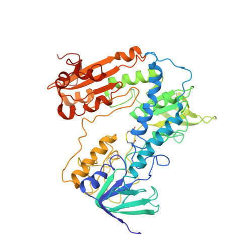
    Trypanothione reductase (TryR) is a genetically validated drug target in the parasite Trypanosoma brucei , the causative agent of human African trypanosomiasis. Here we report the discovery, synthesis, and development of a novel series of TryR inhibitors based on a 3,4-dihydroquinazoline scaffold. In addition, a high resolution crystal structure of TryR, alone and in complex with substrates and inhibitors from this series, is presented. This represents the first report of a high resolution complex between a noncovalent ligand and this enzyme. Structural studies revealed that upon ligand binding the enzyme undergoes a conformational change to create a new subpocket which is occupied by an aryl group on the ligand. Therefore, the inhibitor, in effect, creates its own small binding pocket within the otherwise large, solvent exposed active site. The TryR-ligand structure was subsequently used to guide the synthesis of inhibitors, including analogues that challenged the induced subpocket. This resulted in the development of inhibitors with improved potency against both TryR and T. brucei parasites in a whole cell assay.


  • Organizational Affiliation

    Division of Biological Chemistry and Drug Discovery, College of Life Sciences, University of Dundee , Dow Street, Dundee DD1 5EH, U.K.


Macromolecules
Find similar proteins by:  (by identity cutoff)  |  3D Structure
Entity ID: 1
MoleculeChains Sequence LengthOrganismDetailsImage
TRYPANOTHIONE REDUCTASE
A, B, C, D
495Trypanosoma brucei brucei TREU927Mutation(s): 0 
EC: 1.8.1.12
UniProt
Find proteins for Q389T8 (Trypanosoma brucei brucei (strain 927/4 GUTat10.1))
Explore Q389T8 
Go to UniProtKB:  Q389T8
Entity Groups  
Sequence Clusters30% Identity50% Identity70% Identity90% Identity95% Identity100% Identity
UniProt GroupQ389T8
Sequence Annotations
Expand
  • Reference Sequence
Small Molecules
Ligands 3 Unique
IDChains Name / Formula / InChI Key2D Diagram3D Interactions
FAD
Query on FAD

Download Ideal Coordinates CCD File 
E [auth A],
H [auth B],
K [auth C],
O [auth D]
FLAVIN-ADENINE DINUCLEOTIDE
C27 H33 N9 O15 P2
VWWQXMAJTJZDQX-UYBVJOGSSA-N
WP6
Query on WP6

Download Ideal Coordinates CCD File 
F [auth A],
I [auth B],
L [auth C],
P [auth D]
(4S)-3-BENZYL-6-CHLORO-2-METHYL-4-PHENYL-3,4-DIHYDROQUINAZOLINE
C22 H19 Cl N2
HYYQDTGAVFYWEW-QFIPXVFZSA-N
CL
Query on CL

Download Ideal Coordinates CCD File 
G [auth A],
J [auth B],
M [auth C],
N [auth C],
Q [auth D]
CHLORIDE ION
Cl
VEXZGXHMUGYJMC-UHFFFAOYSA-M
Binding Affinity Annotations 
IDSourceBinding Affinity
WP6 Binding MOAD:  2WP6 IC50: 930 (nM) from 1 assay(s)
BindingDB:  2WP6 IC50: 930 (nM) from 1 assay(s)
Experimental Data & Validation

Experimental Data

  • Method: X-RAY DIFFRACTION
  • Resolution: 2.50 Å
  • R-Value Free: 0.236 
  • R-Value Work: 0.163 
  • R-Value Observed: 0.167 
  • Space Group: P 1 21 1
Unit Cell:
Length ( Å )Angle ( ˚ )
a = 101.65α = 90
b = 63.21β = 97.82
c = 170.5γ = 90
Software Package:
Software NamePurpose
REFMACrefinement
XDSdata reduction
XSCALEdata scaling
MOLREPphasing

Structure Validation

View Full Validation Report


Ligand Structure Quality Assessment 


Entry History 

Deposition Data

Revision History  (Full details and data files)

  • Version 1.0: 2010-10-13
    Type: Initial release
  • Version 1.1: 2011-10-19
    Changes: Database references, Derived calculations, Version format compliance
  • Version 1.2: 2023-12-20
    Changes: Data collection, Database references, Derived calculations, Other, Refinement description