2WOK | pdb_00002wok

Clavulanic acid biosynthesis oligopeptide binding protein 2 complexed with bradykinin


Experimental Data Snapshot

  • Method: X-RAY DIFFRACTION
  • Resolution: 1.70 Å
  • R-Value Free: 
    0.223 (Depositor), 0.221 (DCC) 
  • R-Value Work: 
    0.185 (Depositor), 0.183 (DCC) 
  • R-Value Observed: 
    0.187 (Depositor) 

Starting Model: experimental
View more details

wwPDB Validation 3D Report Full Report

Validation slider image for 2WOK

This is version 1.4 of the entry. See complete history

Literature

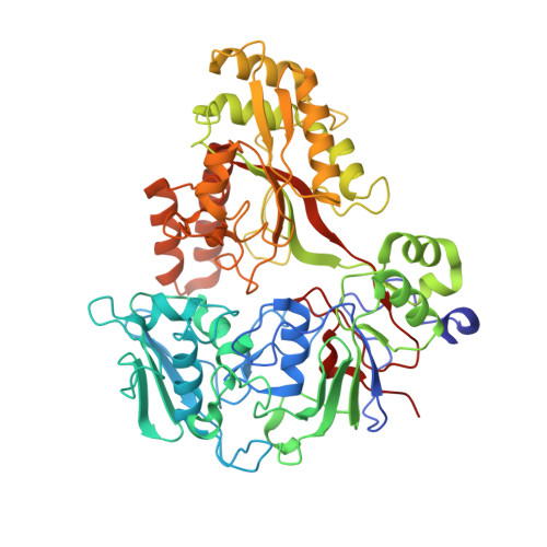
Crystal structures of an oligopeptide-binding protein from the biosynthetic pathway of the beta-lactamase inhibitor clavulanic acid.

Mackenzie, A.K.Valegard, K.Iqbal, A.Caines, M.E.Kershaw, N.J.Jensen, S.E.Schofield, C.J.Andersson, I.

(2010) J Mol Biology 396: 332-344

  • DOI: https://doi.org/10.1016/j.jmb.2009.11.045
  • Primary Citation Related Structures: 
    2WOK, 2WOL, 2WOP

  • PubMed Abstract: 

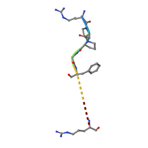
    Clavulanic acid (CA) is a clinically important beta-lactamase inhibitor that is produced by fermentation of Streptomyces clavuligerus. The CA biosynthesis pathway starts from arginine and glyceraldehyde-3-phosphate and proceeds via (3S,5S)-clavaminic acid, which is converted to (3R,5R)-clavaldehyde, the immediate precursor of (3R,5R)-CA. Open reading frames 7 (orf7) and 15 (orf15) of the CA biosynthesis cluster encode oligopeptide-binding proteins (OppA1 and OppA2), which are essential for CA biosynthesis. OppA1/2 are proposed to be involved in the binding and/or transport of peptides across the S. clavuligerus cell membrane. Peptide binding assays reveal that recombinant OppA1 and OppA2 bind di-/tripeptides containing arginine and certain nonapeptides including bradykinin. Crystal structures of OppA2 in its apo form and in complex with arginine or bradykinin were solved to 1.45, 1.7, and 1.7 A resolution, respectively. The overall fold of OppA2 consists of two lobes with a deep cavity in the center, as observed for other oligopeptide-binding proteins. The large cavity creates a peptide/arginine binding cleft. The crystal structures of OppA2 in complex with arginine or bradykinin reveal that the C-terminal arginine of bradykinin binds similarly to arginine. The results are discussed in terms of the possible roles of OppA1/2 in CA biosynthesis.


  • Organizational Affiliation
    • Department of Molecular Biology, Swedish University of Agricultural Sciences, Uppsala, Sweden.

Macromolecule Content 

  • Total Structure Weight: 63.31 kDa 
  • Atom Count: 4,759 
  • Modeled Residue Count: 559 
  • Deposited Residue Count: 571 
  • Unique protein chains: 2

Macromolecules

Find similar proteins by:|  3D Structure
Entity ID: 1
MoleculeChains  Sequence LengthOrganismDetailsImage
CLAVULANIC ACID BIOSYNTHESIS OLIGOPEPTIDE BINDING PROTEIN 2562Streptomyces clavuligerusMutation(s): 0 
UniProt
Find proteins for Q8KRB4 (Streptomyces clavuligerus)
Explore Q8KRB4 
Go to UniProtKB:  Q8KRB4
Entity Groups
Sequence Clusters30% Identity50% Identity70% Identity90% Identity95% Identity100% Identity
UniProt GroupQ8KRB4
Sequence Annotations
Expand
Reference Sequence
Find similar proteins by:  Sequence   |   3D Structure  
Entity ID: 2
MoleculeChains  Sequence LengthOrganismDetailsImage
Kininogen-19Homo sapiensMutation(s): 0 
UniProt & NIH Common Fund Data Resources
Find proteins for P01042 (Homo sapiens)
Explore P01042 
Go to UniProtKB:  P01042
PHAROS:  P01042
GTEx:  ENSG00000113889 
Entity Groups
UniProt GroupP01042
Sequence Annotations
Expand
Reference Sequence

Experimental Data & Validation

Experimental Data

  • Method: X-RAY DIFFRACTION
  • Resolution: 1.70 Å
  • R-Value Free:  0.223 (Depositor), 0.221 (DCC) 
  • R-Value Work:  0.185 (Depositor), 0.183 (DCC) 
  • R-Value Observed: 0.187 (Depositor) 
Space Group: P 1 21 1
Unit Cell:
Length ( Å )Angle ( ˚ )
a = 53.511α = 90
b = 83.327β = 95.54
c = 62.38γ = 90
Software Package:
Software NamePurpose
REFMACrefinement
DENZOdata reduction
SCALEPACKdata scaling
MOLREPphasing

Structure Validation

View Full Validation Report


Entry History 

Deposition Data

Revision History  (Full details and data files)

  • Version 1.0: 2009-12-08
    Type: Initial release
  • Version 1.1: 2011-05-08
    Changes: Version format compliance
  • Version 1.2: 2011-07-13
    Changes: Version format compliance
  • Version 1.3: 2018-12-12
    Changes: Data collection, Database references, Source and taxonomy, Structure summary
  • Version 1.4: 2023-12-20
    Changes: Data collection, Database references, Derived calculations, Other, Refinement description