2WMN

Structure of the complex between DOCK9 and Cdc42-GDP.


Experimental Data Snapshot

  • Method: X-RAY DIFFRACTION
  • Resolution: 2.39 Å
  • R-Value Free: 0.276 
  • R-Value Work: 0.220 
  • R-Value Observed: 0.223 

wwPDB Validation   3D Report Full Report


This is version 1.3 of the entry. See complete history


Literature

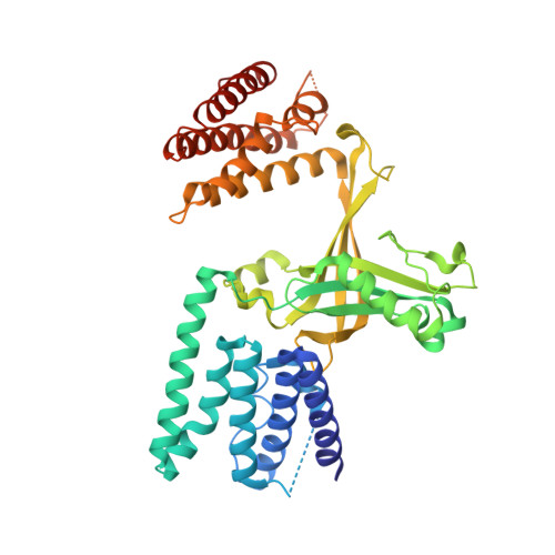
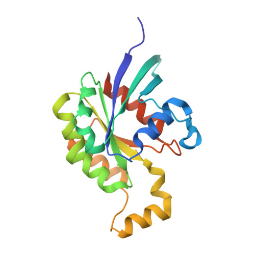
Activation of Rho Gtpases by Dock Exchange Factors is Mediated by a Nucleotide Sensor.

Yang, J.Zhang, Z.Roe, S.M.Marshall, C.J.Barford, D.

(2009) Science 325: 1398

  • DOI: https://doi.org/10.1126/science.1174468
  • Primary Citation of Related Structures:  
    2WM9, 2WMN, 2WMO

  • PubMed Abstract: 

    Activation of Rho guanosine triphosphatases (GTPases) to the guanine triphosphate (GTP)-bound state is a critical event in their regulation of the cytoskeleton and cell signaling. Members of the DOCK family of guanine nucleotide exchange factors (GEFs) are important activators of Rho GTPases, but the mechanism of activation by their catalytic DHR2 domain is unknown. Through structural analysis of DOCK9-Cdc42 complexes, we identify a nucleotide sensor within the alpha10 helix of the DHR2 domain that contributes to release of guanine diphosphate (GDP) and then to discharge of the activated GTP-bound Cdc42. Magnesium exclusion, a critical factor in promoting GDP release, is mediated by a conserved valine residue within this sensor, whereas binding of GTP-Mg2+ to the nucleotide-free complex results in magnesium-inducing displacement of the sensor to stimulate discharge of Cdc42-GTP. These studies identify an unusual mechanism of GDP release and define the complete GEF catalytic cycle from GDP dissociation followed by GTP binding and discharge of the activated GTPase.


  • Organizational Affiliation

    Section of Structural Biology, Institute of Cancer Research, Chester Beatty Laboratories, 237 Fulham Road, London SW3 6JB, UK.


Macromolecules
Find similar proteins by:  (by identity cutoff)  |  3D Structure
Entity ID: 1
MoleculeChains Sequence LengthOrganismDetailsImage
DEDICATOR OF CYTOKINESIS PROTEIN 9428Homo sapiensMutation(s): 0 
UniProt & NIH Common Fund Data Resources
Find proteins for Q9BZ29 (Homo sapiens)
Explore Q9BZ29 
Go to UniProtKB:  Q9BZ29
PHAROS:  Q9BZ29
GTEx:  ENSG00000088387 
Entity Groups  
Sequence Clusters30% Identity50% Identity70% Identity90% Identity95% Identity100% Identity
UniProt GroupQ9BZ29
Sequence Annotations
Expand
  • Reference Sequence
Find similar proteins by:  (by identity cutoff)  |  3D Structure
Entity ID: 2
MoleculeChains Sequence LengthOrganismDetailsImage
CELL DIVISION CONTROL PROTEIN 42 HOMOLOG190Homo sapiensMutation(s): 0 
UniProt & NIH Common Fund Data Resources
Find proteins for P60953 (Homo sapiens)
Explore P60953 
Go to UniProtKB:  P60953
PHAROS:  P60953
GTEx:  ENSG00000070831 
Entity Groups  
Sequence Clusters30% Identity50% Identity70% Identity90% Identity95% Identity100% Identity
UniProt GroupP60953
Sequence Annotations
Expand
  • Reference Sequence
Small Molecules
Ligands 1 Unique
IDChains Name / Formula / InChI Key2D Diagram3D Interactions
GDP
Query on GDP

Download Ideal Coordinates CCD File 
C [auth B]GUANOSINE-5'-DIPHOSPHATE
C10 H15 N5 O11 P2
QGWNDRXFNXRZMB-UUOKFMHZSA-N
Experimental Data & Validation

Experimental Data

  • Method: X-RAY DIFFRACTION
  • Resolution: 2.39 Å
  • R-Value Free: 0.276 
  • R-Value Work: 0.220 
  • R-Value Observed: 0.223 
  • Space Group: P 21 21 2
Unit Cell:
Length ( Å )Angle ( ˚ )
a = 93.05α = 90
b = 88.472β = 90
c = 90.12γ = 90
Software Package:
Software NamePurpose
PHENIXrefinement
iMOSFLMdata reduction
SCALAdata scaling
PHASERphasing

Structure Validation

View Full Validation Report


Entry History 

Deposition Data

Revision History  (Full details and data files)

  • Version 1.0: 2009-09-22
    Type: Initial release
  • Version 1.1: 2011-05-08
    Changes: Version format compliance
  • Version 1.2: 2011-07-13
    Changes: Version format compliance
  • Version 1.3: 2023-12-13
    Changes: Data collection, Database references, Derived calculations, Other, Refinement description