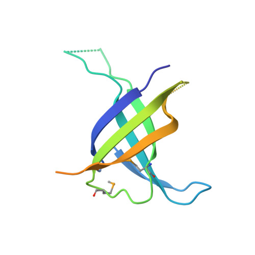
Structure and Function of Phage P2 Orf34(P2), a New Type of Single-Stranded DNA Binding Protein.
Scaltriti, E., Tegoni, M., Rivetti, C., Launay, H., Masson, J.Y., Magadan, A.H., Tremblay, D., Moineau, S., Ramoni, R., Lichiere, J., Campanacci, V., Cambillau, C., Ortiz-Lombardia, M.(2009) Mol Microbiol 73: 1156
- PubMed: 19719513 Search on PubMed
- DOI: https://doi.org/10.1111/j.1365-2958.2009.06844.x
- Primary Citation Related Structures:
2WKC, 2WKD - PubMed Abstract:
Lactococcus lactis, a Gram-positive bacterium widely used by the dairy industry, is subject to infection by a diverse population of virulent phages, predominantly by those of the 936 group, including the siphovirus phage p2. Confronted with the negative impact of phage infection on milk fermentation, the study of the biology of lactococcal provides insight from applied and fundamental perspectives. We decided to characterize the product of the orf34 gene from lactococcus phage p2, which was considered as a candidate single-stranded DNA binding protein (SSB) due to its localization downstream of a gene coding for a single-strand annealing protein. Two-dimensional gel electrophoresis showed that ORF34(p2) is expressed in large amounts during the early phases of phage infection, suggesting an important role in this process. Gel-shift assays, surface plasmon resonance and atomic force microscopy demonstrated that ORF34(p2) interacts with single-strand DNA with nanomolar affinity. We also determined the crystal structure of ORF34(p2) and showed that it bears a variation of the typical oligonucleotide/oligosaccharide binding-fold of SSBs. Finally, we found that ORF34(p2) is able to stimulate Escherichia coli RecA-mediated homologous recombination. The specific structural and biochemical properties that distinguish ORF34(p2) from other SSB proteins are discussed.
- Architecture et Fonction des Macromolécules Biologiques, UMR 6098 CNRS and Universités d'Aix-Marseille I and II, Campus de Luminy, Marseille Cedex 09, France.
Organizational Affiliation: