2WJQ

NanC porin structure in hexagonal crystal form.


Experimental Data Snapshot

  • Method: X-RAY DIFFRACTION
  • Resolution: 2.00 Å
  • R-Value Free: 0.214 
  • R-Value Work: 0.191 
  • R-Value Observed: 0.192 

wwPDB Validation   3D Report Full Report


Ligand Structure Quality Assessment 


This is version 1.3 of the entry. See complete history


Literature

Nanc Crystal Structure, a Model for Outer Membrane Channels of the Acidic Sugar-Specific Kdgm Porin Family.

Wirth, C.Condemine, G.Boiteux, C.Berneche, S.Schirmer, T.Peneff, C.M.

(2009) J Mol Biol 394: 718

  • DOI: https://doi.org/10.1016/j.jmb.2009.09.054
  • Primary Citation of Related Structures:  
    2WJQ, 2WJR

  • PubMed Abstract: 

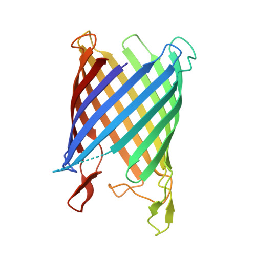
    Sialic acids are acidic sugars present mostly on vertebrate cell surfaces, which can be metabolized by bacteria and act as an inflammation signal. N-Acetylneuraminic acid, the most abundant sialic acid, can enter into Escherichia coli K12 through NanC, an N-acetylneuraminic acid-inducible outer-membrane channel. With its 215 residues, NanC belongs to the family of small monomeric KdgM-related porins. KdgM homologues are found in gammaproteobacteria, including major plant and human pathogens, and together they define a large family of putative acidic sugar/oligosaccharide transporters, which are as yet poorly characterized. Here, we present the first high-resolution structure of a KdgM family member. NanC folds into a 28-A-high, 12-stranded beta-barrel, resembling the beta-domain of autotransporter NalP and defining an open pore with an average radius of 3.3 A. The channel is lined by two strings of basic residues facing each other across the pore, a feature that appears largely conserved within the KdgM family and is likely to facilitate the diffusion of acidic oligosaccharides.


  • Organizational Affiliation

    Department of Structural Biology, Biozentrum, University of Basel, Klingelbergstrasse 70, CH-4056 Basel, Switzerland.


Macromolecules
Find similar proteins by:  (by identity cutoff)  |  3D Structure
Entity ID: 1
MoleculeChains Sequence LengthOrganismDetailsImage
PROBABLE N-ACETYLNEURAMINIC ACID OUTER MEMBRANE CHANNEL PROTEIN NANC215Escherichia coli K-12Mutation(s): 0 
Membrane Entity: Yes 
UniProt
Find proteins for P69856 (Escherichia coli (strain K12))
Explore P69856 
Go to UniProtKB:  P69856
Entity Groups  
Sequence Clusters30% Identity50% Identity70% Identity90% Identity95% Identity100% Identity
UniProt GroupP69856
Sequence Annotations
Expand
  • Reference Sequence
Small Molecules
Ligands 3 Unique
IDChains Name / Formula / InChI Key2D Diagram3D Interactions
LDA
Query on LDA

Download Ideal Coordinates CCD File 
L [auth A],
M [auth A],
N [auth A],
O [auth A]
LAURYL DIMETHYLAMINE-N-OXIDE
C14 H31 N O
SYELZBGXAIXKHU-UHFFFAOYSA-N
OCT
Query on OCT

Download Ideal Coordinates CCD File 
D [auth A]
E [auth A]
F [auth A]
G [auth A]
H [auth A]
D [auth A],
E [auth A],
F [auth A],
G [auth A],
H [auth A],
I [auth A],
J [auth A],
K [auth A],
P [auth A]
N-OCTANE
C8 H18
TVMXDCGIABBOFY-UHFFFAOYSA-N
CL
Query on CL

Download Ideal Coordinates CCD File 
B [auth A],
C [auth A]
CHLORIDE ION
Cl
VEXZGXHMUGYJMC-UHFFFAOYSA-M
Experimental Data & Validation

Experimental Data

  • Method: X-RAY DIFFRACTION
  • Resolution: 2.00 Å
  • R-Value Free: 0.214 
  • R-Value Work: 0.191 
  • R-Value Observed: 0.192 
  • Space Group: P 63 2 2
Unit Cell:
Length ( Å )Angle ( ˚ )
a = 96.823α = 90
b = 96.823β = 90
c = 121.253γ = 120
Software Package:
Software NamePurpose
MOSFLMdata reduction
SCALAdata scaling
SHELXDphasing
SHARPphasing
REFMACrefinement

Structure Validation

View Full Validation Report


Ligand Structure Quality Assessment 


Entry History 

Deposition Data

Revision History  (Full details and data files)

  • Version 1.0: 2009-10-13
    Type: Initial release
  • Version 1.1: 2011-07-13
    Changes: Advisory, Version format compliance
  • Version 1.2: 2019-03-06
    Changes: Data collection, Experimental preparation, Other
  • Version 1.3: 2019-05-08
    Changes: Data collection, Experimental preparation