Structural Basis for Cofactor-Independent Dioxygenation of N-Heteroaromatic Compounds at the {Alpha}/{Beta}-Hydrolase Fold.
Steiner, R.A., Janssen, H.J., Roversi, P., Oakley, A.J., Fetzner, S.(2010) Proc Natl Acad Sci U S A 107: 657
- PubMed: 20080731 Search on PubMedSearch on PubMed Central
- DOI: https://doi.org/10.1073/pnas.0909033107
- Primary Citation Related Structures:
2WJ3, 2WJ4, 2WJ6, 2WM2, 3IBT - PubMed Abstract:
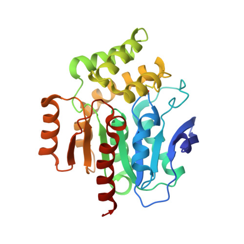
Enzymatic catalysis of oxygenation reactions in the absence of metal or organic cofactors is a considerable biochemical challenge. The CO-forming 1-H-3-hydroxy-4-oxoquinaldine 2,4-dioxygenase (HOD) from Arthrobacter nitroguajacolicus Rü61a and 1-H-3-hydroxy-4-oxoquinoline 2,4-dioxygenase (QDO) from Pseudomonas putida 33/1 are homologous cofactor-independent dioxygenases involved in the breakdown of N-heteroaromatic compounds. To date, they are the only dioxygenases suggested to belong to the alpha/beta-hydrolase fold superfamily. Members of this family typically catalyze hydrolytic processes rather than oxygenation reactions. We present here the crystal structures of both HOD and QDO in their native state as well as the structure of HOD in complex with its natural 1-H-3-hydroxy-4-oxoquinaldine substrate, its N-acetylanthranilate reaction product, and chloride as dioxygen mimic. HOD and QDO are structurally very similar. They possess a classical alpha/beta-hydrolase fold core domain additionally equipped with a cap domain. Organic substrates bind in a preorganized active site with an orientation ideally suited for selective deprotonation of their hydroxyl group by a His/Asp charge-relay system affording the generation of electron-donating species. The "oxyanion hole" of the alpha/beta-hydrolase fold, typically employed to stabilize the tetrahedral intermediate in ester hydrolysis reactions, is utilized here to host and control oxygen chemistry, which is proposed to involve a peroxide anion intermediate. Product release by proton back transfer from the catalytic histidine is driven by minimization of intramolecular charge repulsion. Structural and kinetic data suggest a nonnucleophilic general-base mechanism. Our analysis provides a framework to explain cofactor-independent dioxygenation within a protein architecture generally employed to catalyze hydrolytic reactions.
- Randall Division of Cell and Molecular Biophysics, King's College London, New Hunt's House, Guy's Campus, London SE1 1UL, England. roberto.steiner@kcl.ac.uk
Organizational Affiliation: