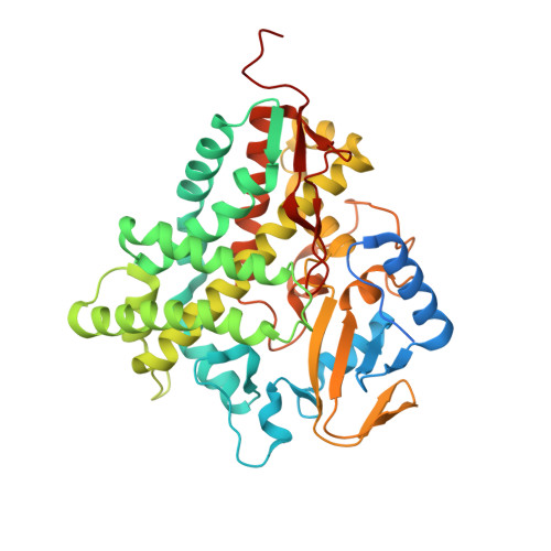
2WIO

Structure of the histidine tagged, open cytochrome P450 Eryk from S. erythraea


Experimental Data Snapshot

  • Method: X-RAY DIFFRACTION
  • Resolution: 2.00 Å
  • R-Value Free: 0.232 
  • R-Value Work: 0.183 
  • R-Value Observed: 0.186 

Starting Model: experimental
View more details

wwPDB Validation   3D Report Full Report


Ligand Structure Quality Assessment 


This is version 1.3 of the entry. See complete history


Literature

Investigating the Structural Plasticity of a Cytochrome P450: Three-Dimensional Structures of P450 Eryk and Binding to its Physiological Substrate.

Savino, C.Montemiglio, L.C.Sciara, G.Miele, A.E.Kendrew, S.G.Jemth, P.Gianni, S.Vallone, B.

(2009) J Biol Chem 284: 29170

  • DOI: https://doi.org/10.1074/jbc.M109.003590
  • Primary Citation of Related Structures:  
    2JJN, 2JJO, 2WIO

  • PubMed Abstract: 

    Cytochrome P450s are heme-containing proteins that catalyze the oxidative metabolism of many physiological endogenous compounds. Because of their unique oxygen chemistry and their key role in drug and xenobiotic metabolism, particular attention has been devoted in elucidating their mechanism of substrate recognition. In this work, we analyzed the three-dimensional structures of a monomeric cytochrome P450 from Saccharopolyspora erythraea, commonly called EryK, and the binding kinetics to its physiological ligand, erythromycin D. Three different structures of EryK were obtained: two ligand-free forms and one in complex with its substrate. Analysis of the substrate-bound structure revealed the key structural determinants involved in substrate recognition and selectivity. Interestingly, the ligand-free structures of EryK suggested that the protein may explore an open and a closed conformation in the absence of substrate. In an effort to validate this hypothesis and to investigate the energetics between such alternative conformations, we performed stopped-flow absorbance experiments. Data demonstrated that EryK binds erythromycin D via a mechanism involving at least two steps. Contrary to previously characterized cytochrome P450s, analysis of double jump mixing experiments confirmed that this complex scenario arises from a pre-existing equilibrium between the open and closed subpopulations of EryK, rather than from an induced-fit type mechanism.


  • Organizational Affiliation

    CNR Institute of Molecular Biology and Pathology, P.le A. Moro 5, 00185 Rome, Italy.


Macromolecules
Find similar proteins by:  (by identity cutoff)  |  3D Structure
Entity ID: 1
MoleculeChains Sequence LengthOrganismDetailsImage
ERYTHROMYCIN B/D C-12 HYDROXYLASE431Saccharopolyspora erythraeaMutation(s): 0 
EC: 1.14
UniProt
Find proteins for P48635 (Saccharopolyspora erythraea (strain ATCC 11635 / DSM 40517 / JCM 4748 / NBRC 13426 / NCIMB 8594 / NRRL 2338))
Explore P48635 
Go to UniProtKB:  P48635
Entity Groups  
Sequence Clusters30% Identity50% Identity70% Identity90% Identity95% Identity100% Identity
UniProt GroupP48635
Sequence Annotations
Expand
  • Reference Sequence
Small Molecules
Ligands 1 Unique
IDChains Name / Formula / InChI Key2D Diagram3D Interactions
HEM
Query on HEM

Download Ideal Coordinates CCD File 
B [auth A]PROTOPORPHYRIN IX CONTAINING FE
C34 H32 Fe N4 O4
KABFMIBPWCXCRK-RGGAHWMASA-L
Experimental Data & Validation

Experimental Data

  • Method: X-RAY DIFFRACTION
  • Resolution: 2.00 Å
  • R-Value Free: 0.232 
  • R-Value Work: 0.183 
  • R-Value Observed: 0.186 
  • Space Group: P 21 21 21
Unit Cell:
Length ( Å )Angle ( ˚ )
a = 37.932α = 90
b = 57.341β = 90
c = 179.557γ = 90
Software Package:
Software NamePurpose
REFMACrefinement
MOSFLMdata reduction
SCALAdata scaling
MOLREPphasing

Structure Validation

View Full Validation Report


Ligand Structure Quality Assessment 


Entry History 

Deposition Data

Revision History  (Full details and data files)

  • Version 1.0: 2009-07-21
    Type: Initial release
  • Version 1.1: 2011-05-08
    Changes: Version format compliance
  • Version 1.2: 2011-07-13
    Changes: Version format compliance
  • Version 1.3: 2023-12-13
    Changes: Data collection, Database references, Derived calculations, Other, Refinement description