2WFV

Crystal structure of DILP5 variant C4


Experimental Data Snapshot

  • Method: X-RAY DIFFRACTION
  • Resolution: 1.85 Å
  • R-Value Free: 0.232 
  • R-Value Work: 0.199 
  • R-Value Observed: 0.202 

wwPDB Validation   3D Report Full Report


This is version 1.2 of the entry. See complete history


Literature

Structural and Biological Properties of the Drosophila Insulin-Like Peptide 5 Show Evolutionary Conservation.

Sajid, W.Kulahin, N.Schluckebier, G.Ribel, U.Henderson, H.R.Tatar, M.Hansen, B.F.Svendsen, A.M.Kiselyov, V.V.Norgaard, P.Wahlund, P.Brandt, J.Kohanski, R.A.Andersen, A.S.De Meyts, P.

(2011) J Biol Chem 286: 661

  • DOI: https://doi.org/10.1074/jbc.M110.156018
  • Primary Citation of Related Structures:  
    2WFU, 2WFV

  • PubMed Abstract: 

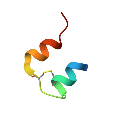
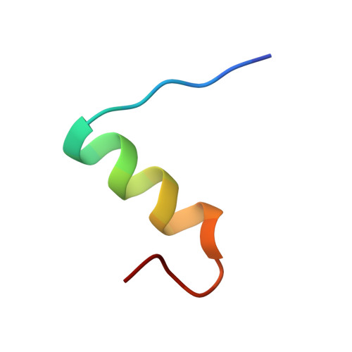
    We report the crystal structure of two variants of Drosophila melanogaster insulin-like peptide 5 (DILP5) at a resolution of 1.85 Å. DILP5 shares the basic fold of the insulin peptide family (T conformation) but with a disordered B-chain C terminus. DILP5 dimerizes in the crystal and in solution. The dimer interface is not similar to that observed in vertebrates, i.e. through an anti-parallel β-sheet involving the B-chain C termini but, in contrast, is formed through an anti-parallel β-sheet involving the B-chain N termini. DILP5 binds to and activates the human insulin receptor and lowers blood glucose in rats. It also lowers trehalose levels in Drosophila. Reciprocally, human insulin binds to the Drosophila insulin receptor and induces negative cooperativity as in the human receptor. DILP5 also binds to insect insulin-binding proteins. These results show high evolutionary conservation of the insulin receptor binding properties despite divergent insulin dimerization mechanisms.


  • Organizational Affiliation

    Receptor Systems Biology Laboratory, Insulin and Incretin Biology, Hagedorn Research Institute, 2820 Gentofte, Denmark.


Macromolecules

Find similar proteins by:  Sequence   |   3D Structure  

Entity ID: 1
MoleculeChains Sequence LengthOrganismDetailsImage
PROBABLE INSULIN-LIKE PEPTIDE 5 A CHAIN25Drosophila melanogasterMutation(s): 1 
UniProt
Find proteins for Q7KUD5 (Drosophila melanogaster)
Explore Q7KUD5 
Go to UniProtKB:  Q7KUD5
Entity Groups  
Sequence Clusters30% Identity50% Identity70% Identity90% Identity95% Identity100% Identity
UniProt GroupQ7KUD5
Sequence Annotations
Expand
  • Reference Sequence

Find similar proteins by:  Sequence   |   3D Structure  

Entity ID: 2
MoleculeChains Sequence LengthOrganismDetailsImage
PROBABLE INSULIN-LIKE PEPTIDE 5 B CHAIN23Drosophila melanogasterMutation(s): 0 
UniProt
Find proteins for Q7KUD5 (Drosophila melanogaster)
Explore Q7KUD5 
Go to UniProtKB:  Q7KUD5
Entity Groups  
Sequence Clusters30% Identity50% Identity70% Identity90% Identity95% Identity100% Identity
UniProt GroupQ7KUD5
Sequence Annotations
Expand
  • Reference Sequence
Experimental Data & Validation

Experimental Data

  • Method: X-RAY DIFFRACTION
  • Resolution: 1.85 Å
  • R-Value Free: 0.232 
  • R-Value Work: 0.199 
  • R-Value Observed: 0.202 
  • Space Group: P 43 21 2
Unit Cell:
Length ( Å )Angle ( ˚ )
a = 40.445α = 90
b = 40.445β = 90
c = 45.241γ = 90
Software Package:
Software NamePurpose
REFMACrefinement
HKL-2000data reduction
HKL-2000data scaling
SHARPphasing

Structure Validation

View Full Validation Report


Entry History 

Deposition Data

Revision History  (Full details and data files)

  • Version 1.0: 2010-05-26
    Type: Initial release
  • Version 1.1: 2011-07-13
    Changes: Advisory, Version format compliance
  • Version 1.2: 2012-04-18
    Changes: Other