2W9Y | pdb_00002w9y

The structure of the lipid binding protein Ce-FAR-7 from Caenorhabditis elegans


Experimental Data Snapshot

  • Method: X-RAY DIFFRACTION
  • Resolution: 1.80 Å
  • R-Value Free: 
    0.251 (Depositor), 0.242 (DCC) 
  • R-Value Work: 
    0.171 (Depositor), 0.167 (DCC) 
  • R-Value Observed: 
    0.174 (Depositor) 

wwPDB Validation 3D Report Full Report

Validation slider image for 2W9Y

This is version 1.2 of the entry. See complete history

Literature

Fatty Acid and Retinoid Binding Proteins Have Distinct Binding Pockets for the Two Types of Cargo

Jordanova, R.Groves, M.R.Kostova, E.B.Woltersdorf, C.Liebau, E.Tucker, P.A.

(2009) J Biological Chem 284: 35818

  • DOI: https://doi.org/10.1074/jbc.M109.022731
  • Primary Citation Related Structures: 
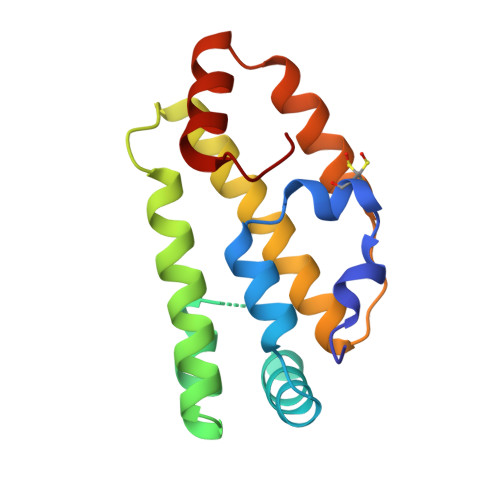
    2W9Y

  • PubMed Abstract: 

    Parasitic nematodes cause serious diseases in humans, animals, and plants. They have limited lipid metabolism and are reliant on lipid-binding proteins to acquire these metabolites from their hosts. Several structurally novel families of lipid-binding proteins in nematodes have been described, including the fatty acid- and retinoid-binding protein family (FAR). In Caenorhabditis elegans, used as a model for studying parasitic nematodes, eight C. elegans FAR proteins have been described. The crystal structure of C. elegans FAR-7 is the first structure of a FAR protein, and it exhibits a novel fold. It differs radically from the mammalian fatty acid-binding proteins and has two ligand binding pockets joined by a surface groove. The first can accommodate the aliphatic chain of fatty acids, whereas the second can accommodate the bulkier retinoids. In addition to demonstrating lipid binding by fluorescence spectroscopy, we present evidence that retinol binding is positively regulated by casein kinase II phosphorylation at a conserved site near the bottom of the second pocket. far-7::GFP (green fluorescent protein) expression shows that it is localized in the head hypodermal syncytia and the excretory cell but that this localization changes under starvation conditions. In conclusion, our study provides the basic structural and functional information for investigation of inhibitors of lipid binding by FAR proteins.


  • Organizational Affiliation
    • European Molecular Biology Laboratory, Hamburg Outstation, 22603 Hamburg, Germany.

Macromolecule Content 

  • Total Structure Weight: 15.73 kDa 
  • Atom Count: 1,278 
  • Modeled Residue Count: 136 
  • Deposited Residue Count: 140 
  • Unique protein chains: 1

Macromolecules

Find similar proteins by:|  3D Structure
Entity ID: 1
MoleculeChains  Sequence LengthOrganismDetailsImage
FATTY ACID/RETINOL BINDING PROTEIN PROTEIN 7, ISOFORM A, CONFIRMED BY TRANSCRIPT EVIDENCE140Caenorhabditis elegansMutation(s): 0 
UniProt
Find proteins for Q9TZ51 (Caenorhabditis elegans)
Explore Q9TZ51 
Go to UniProtKB:  Q9TZ51
Entity Groups
Sequence Clusters30% Identity50% Identity70% Identity90% Identity95% Identity100% Identity
UniProt GroupQ9TZ51
Sequence Annotations
Expand
Reference Sequence

Small Molecules

Modified Residues  1 Unique
IDChains TypeFormula2D DiagramParent
CSX
Query on CSX
A
L-PEPTIDE LINKINGC3 H7 N O3 SCYS

Experimental Data & Validation

Experimental Data

  • Method: X-RAY DIFFRACTION
  • Resolution: 1.80 Å
  • R-Value Free:  0.251 (Depositor), 0.242 (DCC) 
  • R-Value Work:  0.171 (Depositor), 0.167 (DCC) 
  • R-Value Observed: 0.174 (Depositor) 
Space Group: P 21 21 21
Unit Cell:
Length ( Å )Angle ( ˚ )
a = 33.85α = 90
b = 41.6β = 90
c = 101.74γ = 90
Software Package:
Software NamePurpose
REFMACrefinement
XDSdata reduction
SCALAdata scaling
autoSHARPphasing

Structure Validation

View Full Validation Report


Entry History 

Deposition Data

Revision History  (Full details and data files)

  • Version 1.0: 2009-10-20
    Type: Initial release
  • Version 1.1: 2015-04-29
    Changes: Data collection, Derived calculations, Other, Source and taxonomy, Version format compliance
  • Version 1.2: 2025-04-09
    Changes: Data collection, Database references, Derived calculations, Other, Structure summary