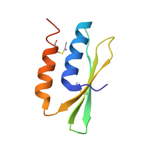
Structure of Srp14 from the Schizosaccharomyces Pombe Signal Recognition Particle.
Brooks, M.A., Ravelli, R.B.G., Mccarthy, A.A., Strub, K., Cusack, S.(2009) Acta Crystallogr D Biol Crystallogr 65: 421
- PubMed: 19390147 Search on PubMed
- DOI: https://doi.org/10.1107/S0907444909005484
- Primary Citation Related Structures:
2W9J - PubMed Abstract:
The signal recognition particle (SRP) Alu domain has been implicated in translation elongation arrest in yeasts and mammals. Fission yeast SRP RNA is similar to that of mammals, but has a minimal Alu-domain RNA lacking two stem-loops. The mammalian Alu-domain proteins SRP9 and SRP14 bind their cognate Alu RNA as a heterodimer. However, in yeasts, notably Saccharomyces cerevisiae, SRP14 is thought to bind Alu RNA as a homodimer, the SRP9 protein being replaced by SRP21, the function of which is not yet clear. Structural characterization of the Schizosaccharomyces pombe Alu domain may thus help to identify the critical features required for elongation arrest. Here, the crystal structure of the SRP14 subunit of S. pombe SRP (SpSRP14) which crystallizes as a homodimer, is presented. Comparison of the SpSRP14 homodimer with the known structure of human SRP9/14 in complex with Alu RNA suggests that many of the protein-RNA contacts centred on the conserved U-turn motif are likely to be conserved in fission yeast. Initial attempts to solve the structure using traditional selenomethionine SAD labelling failed. However, two As atoms originating from the cacodylate buffer were found to make cysteine adducts and strongly contributed to the anomalous substructure. These adducts were highly radiation-sensitive and this property was exploited using the RIP (radiation-damage-induced phasing) method. The combination of SAD and RIP phases yielded an interpretable electron-density map. This example will be of general interest to crystallographers attempting de novo phasing from crystals grown in cacodylate buffer.
- IBBMC-CNRS UMR8619, Bâtiment 430, Université de Paris-Sud, Orsay, France.
Organizational Affiliation: