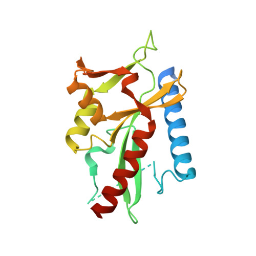
2W8M | pdb_00002w8m

Structure of D212, a nuclease from a fusselovirus.


Experimental Data Snapshot

  • Method: X-RAY DIFFRACTION
  • Resolution: 2.40 Å
  • R-Value Free: 
    0.245 (Depositor), 0.263 (DCC) 
  • R-Value Work: 
    0.209 (Depositor), 0.215 (DCC) 
  • R-Value Observed: 
    0.211 (Depositor) 

wwPDB Validation 3D Report Full Report

Validation slider image for 2W8M

This is version 1.3 of the entry. See complete history

Literature

The crystal structure of D212 from sulfolobus spindle-shaped virus ragged hills reveals a new member of the PD-(D/E)XK nuclease superfamily.

Menon, S.K.Eilers, B.J.Young, M.J.Lawrence, C.M.

(2010) J Virol 84: 5890-5897

  • DOI: https://doi.org/10.1128/JVI.01663-09
  • Primary Citation Related Structures: 
    2W8M

  • PubMed Abstract: 

    Structural studies have made significant contributions to our understanding of Sulfolobus spindle-shaped viruses (Fuselloviridae), an important model system for archaeal viruses. Continuing these efforts, we report the structure of D212 from Sulfolobus spindle-shaped virus Ragged Hills. The overall fold and conservation of active site residues place D212 in the PD-(D/E)XK nuclease superfamily. The greatest structural similarity is found to the archaeal Holliday junction cleavage enzymes, strongly suggesting a role in DNA replication, repair, or recombination. Other roles associated with nuclease activity are also considered.


  • Organizational Affiliation
    • Thermal Biology Institute, Montana State University, Bozeman, MT 59717, USA.

Macromolecule Content 

  • Total Structure Weight: 50.21 kDa 
  • Atom Count: 2,892 
  • Modeled Residue Count: 333 
  • Deposited Residue Count: 424 
  • Unique protein chains: 1

Macromolecules

Find similar proteins by:|  3D Structure
Entity ID: 1
MoleculeChains  Sequence LengthOrganismDetailsImage
ORF D212
A, B
212Sulfolobus virus Ragged HillsMutation(s): 0 
UniProt
Find proteins for Q6TRV5 (Sulfolobus virus Ragged Hills)
Explore Q6TRV5 
Go to UniProtKB:  Q6TRV5
Entity Groups
Sequence Clusters30% Identity50% Identity70% Identity90% Identity95% Identity100% Identity
UniProt GroupQ6TRV5
Sequence Annotations
Expand
Reference Sequence

Experimental Data & Validation

Experimental Data

  • Method: X-RAY DIFFRACTION
  • Resolution: 2.40 Å
  • R-Value Free:  0.245 (Depositor), 0.263 (DCC) 
  • R-Value Work:  0.209 (Depositor), 0.215 (DCC) 
  • R-Value Observed: 0.211 (Depositor) 
Space Group: P 1 21 1
Unit Cell:
Length ( Å )Angle ( ˚ )
a = 56.477α = 90
b = 81.971β = 115.4
c = 60.16γ = 90
Software Package:
Software NamePurpose
DENZOdata reduction
SCALEPACKdata scaling
SOLVEphasing
RESOLVEphasing
REFMACrefinement

Structure Validation

View Full Validation Report


Entry History 

Deposition Data

Revision History  (Full details and data files)

  • Version 1.0: 2009-02-03
    Type: Initial release
  • Version 1.1: 2011-07-13
    Changes: Advisory, Refinement description, Version format compliance
  • Version 1.2: 2018-06-13
    Changes: Advisory, Data collection, Database references
  • Version 1.3: 2024-05-08
    Changes: Advisory, Data collection, Database references, Other