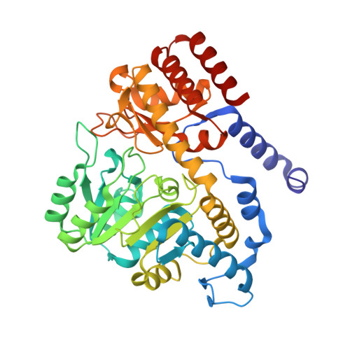
Importance of Tyrosine Residues of Bacillus Stearothermophilus Serine Hydroxymethyltransferase in Cofactor Binding and L-Allo-Thr Cleavage.
Bhavani, B.S., Rajaram, V., Bisht, S., Kaul, P., Prakash, V., Murthy, M.R.N., Appaji Rao, N., Savithri, H.S.(2008) FEBS J 275: 4606
- PubMed: 18699779
- DOI: https://doi.org/10.1111/j.1742-4658.2008.06603.x
- Primary Citation of Related Structures:
2W7D, 2W7E, 2W7F, 2W7G, 2W7H, 2W7I, 2W7J, 2W7K, 2W7L, 2W7M - PubMed Abstract:
Serine hydroxymethyltransferase (SHMT) from Bacillus stearothermophilus (bsSHMT) is a pyridoxal 5'-phosphate-dependent enzyme that catalyses the conversion of L-serine and tetrahydrofolate to glycine and 5,10-methylene tetrahydrofolate. In addition, the enzyme catalyses the tetrahydrofolate-independent cleavage of 3-hydroxy amino acids and transamination. In this article, we have examined the mechanism of the tetrahydrofolate-independent cleavage of 3-hydroxy amino acids by SHMT. The three-dimensional structure and biochemical properties of Y51F and Y61A bsSHMTs and their complexes with substrates, especially L-allo-Thr, show that the cleavage of 3-hydroxy amino acids could proceed via Calpha proton abstraction rather than hydroxyl proton removal. Both mutations result in a complete loss of tetrahydrofolate-dependent and tetrahydrofolate-independent activities. The mutation of Y51 to F strongly affects the binding of pyridoxal 5'-phosphate, possibly as a consequence of a change in the orientation of the phenyl ring in Y51F bsSHMT. The mutant enzyme could be completely reconstituted with pyridoxal 5'-phosphate. However, there was an alteration in the lambda max value of the internal aldimine (396 nm), a decrease in the rate of reduction with NaCNBH3 and a loss of the intermediate in the interaction with methoxyamine (MA). The mutation of Y61 to A results in the loss of interaction with Calpha and Cbeta of the substrates. X-Ray structure and visible CD studies show that the mutant is capable of forming an external aldimine. However, the formation of the quinonoid intermediate is hindered. It is suggested that Y61 is involved in the abstraction of the Calpha proton from 3-hydroxy amino acids. A new mechanism for the cleavage of 3-hydroxy amino acids via Calpha proton abstraction by SHMT is proposed.
- Protein Chemistry and Technology, Central Food Technological Research Institute, Mysore, India.
Organizational Affiliation:


