2W43 | pdb_00002w43

Structure of L-haloacid dehalogenase from S. tokodaii


Experimental Data Snapshot

  • Method: X-RAY DIFFRACTION
  • Resolution: 1.66 Å
  • R-Value Free: 
    0.221 (Depositor), 0.244 (DCC) 
  • R-Value Work: 
    0.177 (Depositor) 
  • R-Value Observed: 
    0.178 (Depositor) 

Starting Model: experimental
View more details

wwPDB Validation 3D Report Full Report

Validation slider image for 2W43

This is version 1.4 of the entry. See complete history

Literature

Biochemical and Structural Studies of a L-Haloacid Dehalogenase from the Thermophilic Archaeon Sulfolobus Tokodaii.

Rye, C.A.Isupov, M.N.Lebedev, A.A.Littlechild, J.A.

(2009) Extremophiles 13: 179

  • DOI: https://doi.org/10.1007/s00792-008-0208-0
  • Primary Citation Related Structures: 
    2W11, 2W43

  • PubMed Abstract: 

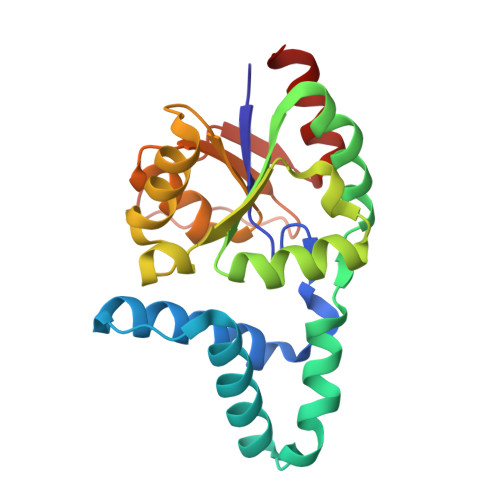
    Haloacid dehalogenases have potential applications in the pharmaceutical and fine chemical industry as well as in the remediation of contaminated land. The L: -2-haloacid dehalogenase from the thermophilic archaeon Sulfolobus tokodaii has been cloned and over-expressed in Escherichia coli and successfully purified to homogeneity. Here we report the structure of the recombinant dehalogenase solved by molecular replacement in two different crystal forms. The enzyme is a homodimer with each monomer being composed of a core-domain of a beta-sheet bundle surrounded by alpha-helices and an alpha-helical sub-domain. This fold is similar to previously solved mesophilic L: -haloacid dehalogenase structures. The monoclinic crystal form contains a putative inhibitor L: -lactate in the active site. The enzyme displays haloacid dehalogenase activity towards carboxylic acids with the halide attached at the C2 position with the highest activity towards chloropropionic acid. The enzyme is thermostable with maximum activity at 60 degrees C and a half-life of over 1 h at 70 degrees C. The enzyme is relatively stable to solvents with 25% activity lost when incubated for 1 h in 20% v/v DMSO.


  • Organizational Affiliation
    • Henry Wellcome Building for Biocatalysis, School of Biosciences, University of Exeter, Stocker Road, Exeter EX4 4QD, UK.

Macromolecule Content 

  • Total Structure Weight: 47.66 kDa 
  • Atom Count: 4,167 
  • Modeled Residue Count: 401 
  • Deposited Residue Count: 402 
  • Unique protein chains: 1

Macromolecules

Find similar proteins by:|  3D Structure
Entity ID: 1
MoleculeChains  Sequence LengthOrganismDetailsImage
HYPOTHETICAL 2-HALOALKANOIC ACID DEHALOGENASE
A, B
201Sulfurisphaera tokodaiiMutation(s): 0 
EC: 3.1.8.2 (PDB Primary Data), 3.8.1.2 (UniProt)
UniProt
Find proteins for Q96XE7 (Sulfurisphaera tokodaii (strain DSM 16993 / JCM 10545 / NBRC 100140 / 7))
Explore Q96XE7 
Go to UniProtKB:  Q96XE7
Entity Groups
Sequence Clusters30% Identity50% Identity70% Identity90% Identity95% Identity100% Identity
UniProt GroupQ96XE7
Sequence Annotations
Expand
Reference Sequence

Experimental Data & Validation

Experimental Data

  • Method: X-RAY DIFFRACTION
  • Resolution: 1.66 Å
  • R-Value Free:  0.221 (Depositor), 0.244 (DCC) 
  • R-Value Work:  0.177 (Depositor) 
  • R-Value Observed: 0.178 (Depositor) 
Space Group: P 21 21 21
Unit Cell:
Length ( Å )Angle ( ˚ )
a = 69.796α = 90
b = 71.889β = 90
c = 85.601γ = 90
Software Package:
Software NamePurpose
REFMACrefinement
DENZOdata reduction
SCALEPACKdata scaling
MOLREPphasing

Structure Validation

View Full Validation Report


Entry History 

Deposition Data

Revision History  (Full details and data files)

  • Version 1.0: 2008-12-09
    Type: Initial release
  • Version 1.1: 2011-05-08
    Changes: Version format compliance
  • Version 1.2: 2011-07-13
    Changes: Version format compliance
  • Version 1.3: 2018-05-30
    Changes: Data collection
  • Version 1.4: 2023-12-13
    Changes: Data collection, Database references, Derived calculations, Other, Refinement description