2W2P | pdb_00002w2p

PCSK9-deltaC D374A mutant bound to WT EGF-A of LDLR


Experimental Data Snapshot

  • Method: X-RAY DIFFRACTION
  • Resolution: 2.62 Å
  • R-Value Free: 
    0.270 (Depositor), 0.268 (DCC) 
  • R-Value Work: 
    0.226 (Depositor), 0.225 (DCC) 
  • R-Value Observed: 
    0.228 (Depositor) 

Starting Model: experimental
View more details

wwPDB Validation 3D Report Full Report

Validation slider image for 2W2P

This is version 1.3 of the entry. See complete history

Literature

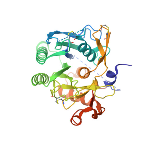
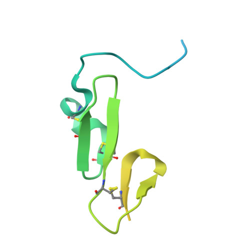
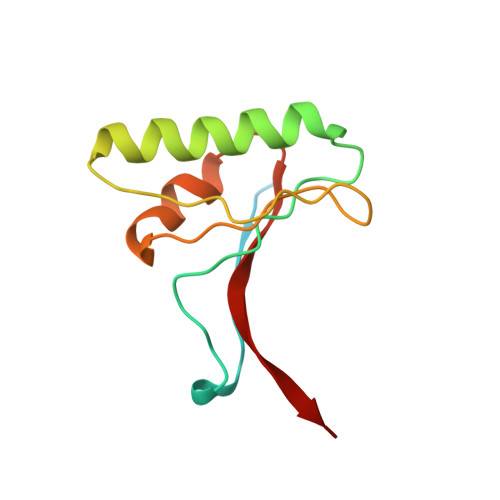
Structural and Biochemical Characterization of the Wild Type Pcsk9/Egf-Ab Complex and Natural Fh Mutants.

Bottomley, M.J.Cirillo, A.Orsatti, L.Ruggeri, L.Fisher, T.S.Santoro, J.C.Cummings, R.T.Cubbon, R.M.Lo Surdo, P.Calzetta, A.Noto, A.Baysarowich, J.Mattu, M.Talamo, F.De Francesco, R.Sparrow, C.P.Sitlani, A.Carfi, A.

(2009) J Biological Chem 284: 1313

  • DOI: https://doi.org/10.1074/jbc.M808363200
  • Primary Citation Related Structures: 
    2W2M, 2W2N, 2W2O, 2W2P, 2W2Q

  • PubMed Abstract: 

    PCSK9 regulates low density lipoprotein receptor (LDLR) levels and consequently is a target for the prevention of atherosclerosis and coronary heart disease. Here we studied the interaction, of LDLR EGF(A/AB) repeats with PCSK9. We show that PCSK9 binds the EGF(AB) repeats in a pH-dependent manner. Although the PCSK9 C-terminal domain is not involved in LDLR binding, PCSK9 autocleavage is required. Moreover, we report the x-ray structure of the PCSK9DeltaC-EGF(AB) complex at neutral pH. Compared with the low pH PCSK9-EGF(A) structure, the new structure revealed rearrangement of the EGF(A) His-306 side chain and disruption of the salt bridge with PCSK9 Asp-374, thus suggesting the basis for enhanced interaction at low pH. In addition, the structure of PCSK9DeltaC bound to EGF(AB)(H306Y), a mutant associated with familial hypercholesterolemia (FH), reveals that the Tyr-306 side chain forms a hydrogen bond with PCSK9 Asp-374, thus mimicking His-306 in the low pH conformation. Consistently, Tyr-306 confers increased affinity for PCSK9. Importantly, we found that although the EGF(AB)(H306Y)-PCSK9 interaction is pH-independent, LDLR(H306Y) binds PCSK9 50-fold better at low pH, suggesting that factors other than His-306 contribute to the pH dependence of PCSK9-LDLR binding. Further, we determined the structures of EGF(AB) bound to PCSK9DeltaC containing the FH-associated D374Y and D374H mutations, revealing additional interactions with EGF(A) mediated by Tyr-374/His-374 and providing a rationale for their disease phenotypes. Finally, we report the inhibitory properties of EGF repeats in a cellular assay measuring LDL uptake.


  • Organizational Affiliation
    • Department of Biochemistry, Istituto di Ricerca di Biologia Molecolare "P. Angeletti", Via Pontina Km 30.600, 00040 Pomezia (Rome), Italy. matthew_bottomley@merck.com

Macromolecule Content 

  • Total Structure Weight: 57.88 kDa 
  • Atom Count: 3,196 
  • Modeled Residue Count: 415 
  • Deposited Residue Count: 533 
  • Unique protein chains: 3

Macromolecules

Find similar proteins by:|  3D Structure
Entity ID: 1
MoleculeChains  Sequence LengthOrganismDetailsImage
PROPROTEIN CONVERTASE SUBTILISIN/KEXIN TYPE 9312Homo sapiensMutation(s): 1 
EC: 3.4.21
UniProt & NIH Common Fund Data Resources
Find proteins for Q8NBP7 (Homo sapiens)
Explore Q8NBP7 
Go to UniProtKB:  Q8NBP7
PHAROS:  Q8NBP7
GTEx:  ENSG00000169174 
Entity Groups
Sequence Clusters30% Identity50% Identity70% Identity90% Identity95% Identity100% Identity
UniProt GroupQ8NBP7
Sequence Annotations
Expand
Reference Sequence
Find similar proteins by:|  3D Structure
Entity ID: 2
MoleculeChains  Sequence LengthOrganismDetailsImage
LOW-DENSITY LIPOPROTEIN RECEPTORB [auth E]107Homo sapiensMutation(s): 0 
UniProt & NIH Common Fund Data Resources
Find proteins for P01130 (Homo sapiens)
Explore P01130 
Go to UniProtKB:  P01130
PHAROS:  P01130
GTEx:  ENSG00000130164 
Entity Groups
Sequence Clusters30% Identity50% Identity70% Identity90% Identity95% Identity100% Identity
UniProt GroupP01130
Sequence Annotations
Expand
Reference Sequence
Find similar proteins by:|  3D Structure
Entity ID: 3
MoleculeChains  Sequence LengthOrganismDetailsImage
PROPROTEIN CONVERTASE SUBTILISIN/KEXIN TYPE 9C [auth P]114Homo sapiensMutation(s): 0 
EC: 3.4.21
UniProt & NIH Common Fund Data Resources
Find proteins for Q8NBP7 (Homo sapiens)
Explore Q8NBP7 
Go to UniProtKB:  Q8NBP7
PHAROS:  Q8NBP7
GTEx:  ENSG00000169174 
Entity Groups
Sequence Clusters30% Identity50% Identity70% Identity90% Identity95% Identity100% Identity
UniProt GroupQ8NBP7
Sequence Annotations
Expand
Reference Sequence

Small Molecules

Ligands 1 Unique
IDChains Name / Formula / InChI Key2D Diagram3D Interactions
CA

Query on CA


Download:Ideal Coordinates CCD File
D [auth E]CALCIUM ION
Ca
BHPQYMZQTOCNFJ-UHFFFAOYSA-N

Experimental Data & Validation

Experimental Data

  • Method: X-RAY DIFFRACTION
  • Resolution: 2.62 Å
  • R-Value Free:  0.270 (Depositor), 0.268 (DCC) 
  • R-Value Work:  0.226 (Depositor), 0.225 (DCC) 
  • R-Value Observed: 0.228 (Depositor) 
Space Group: P 43 21 2
Unit Cell:
Length ( Å )Angle ( ˚ )
a = 84.106α = 90
b = 84.106β = 90
c = 209.745γ = 90
Software Package:
Software NamePurpose
REFMACrefinement
MOSFLMdata reduction
SCALAdata scaling
PHASERphasing

Structure Validation

View Full Validation Report


Entry History 

Revision History  (Full details and data files)

  • Version 1.0: 2008-11-18
    Type: Initial release
  • Version 1.1: 2011-07-13
    Changes: Advisory, Version format compliance
  • Version 1.2: 2023-12-13
    Changes: Data collection, Database references, Derived calculations, Other, Refinement description
  • Version 1.3: 2024-11-13
    Changes: Structure summary