Ligand-Induced Structural Transitions, Mutational Analysis, and 'Open' Quaternary Structure of the M. Tuberculosis Feast/Famine Regulatory Protein (Rv3291C).
Shrivastava, T., Dey, A., Ramachandran, R.(2009) J Mol Biol 392: 1007
- PubMed: 19651141 Search on PubMed
- DOI: https://doi.org/10.1016/j.jmb.2009.07.084
- Primary Citation Related Structures:
2W24, 2W25, 2W29 - PubMed Abstract:
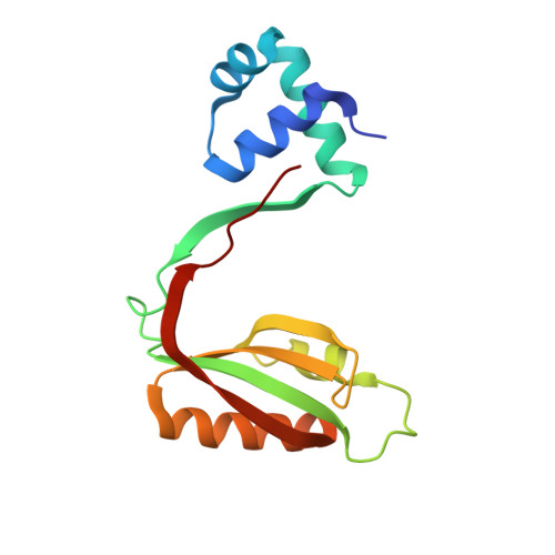
Rv3291c is a member of the feast/famine regulatory protein family that is known to form stable protein-DNA complexes. We report a specific oligomeric transition between hexadecameric and octameric/lower-order oligomers in the presence of Phe that supports an effector-mediated model for the disassembly of a nucleosome-like particle. We had generated two mutants, Gly102Thr and Glu104Ala, which are part of the essential 100-106 effector-binding loop. The Gly102Thr mutant adopts an unusual 'open' quaternary structure and offers interesting functional insights co-related to the binding of an effector. This is similar to the previously reported Escherichia coli Lrp co-crystallized in the presence of DNA where the interactions of the substrate with the N-terminal DNA binding domain presumably lead to symmetry deviations to the oligomeric association. The present structure represents a direct evidence to support that changes made to the effector-binding domain at the C-terminus also result in a functionally relevant quaternary structural change. Conversely, the Glu104Ala mutant retains the closed quaternary association observed in the native protein and reveals nonsymmetrical interaction effects in the two subunits of the dimer. We also report that the native protein unexpectedly binds Lys but does not recognize Arg and offer a structural explanation for it. Error-scaled difference distance matrix analysis suggests that the protein has a relatively flexible core that is presumably needed to mediate the structural changes necessary for the protein's regulatory functions.
- Molecular and Structural Biology Division, Central Drug Research Institute, P.O. Box 173, Chattar Manzil, Mahatma Gandhi Marg, Lucknow 226001, India.
Organizational Affiliation: