2W0D | pdb_00002w0d

Does a Fast Nuclear Magnetic Resonance Spectroscopy- and X-Ray Crystallography Hybrid Approach Provide Reliable Structural Information of Ligand-Protein Complexes? A Case Study of Metalloproteinases.


Experimental Data Snapshot

  • Method: X-RAY DIFFRACTION
  • Resolution: 2.00 Å
  • R-Value Free: 
    0.237 (Depositor), 0.239 (DCC) 
  • R-Value Work: 
    0.179 (Depositor), 0.181 (DCC) 
  • R-Value Observed: 
    0.182 (Depositor) 

Starting Model: other
View more details

wwPDB Validation 3D Report Full Report

Validation slider image for 2W0D

Ligand Structure Quality Assessment 


This is version 1.4 of the entry. See complete history

Literature

Does a Fast Nuclear Magnetic Resonance Spectroscopy- and X-Ray Crystallography Hybrid Approach Provide Reliable Structural Information of Ligand-Protein Complexes? a Case Study of Metalloproteinases.

Isaksson, J.Nystrom, S.Derbyshire, D.J.Wallberg, H.Agback, T.Kovacs, H.Bertini, I.Felli, I.C.

(2009) J Med Chem 52: 1712

  • DOI: https://doi.org/10.1021/jm801388q
  • Primary Citation Related Structures: 
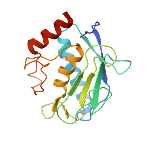
    2W0D

  • PubMed Abstract: 

    A human matrix metalloproteinase (MMP) hydroxamic acid inhibitor (CGS27023A) was cross-docked into 15 MMP-12, MMP-13, MMP-9, and MMP-1 cocrystal structures. The aim was to validate a fast protocol for ligand binding conformation elucidation and to probe the feasibility of using inhibitor-protein NMR contacts to dock an inhibitor into related MMP crystal structures. Such an approach avoids full NMR structure elucidation, saving both spectrometer- and analysis time. We report here that for the studied MMPs, one can obtain docking results well within 1 A compared to the corresponding reference X-ray structure, using backbone amide contacts only. From the perspective of the pharmaceutical industry, these results are relevant for the binding studies of inhibitor series to a common target and have the potential advantage of obtaining information on protein-inhibitor complexes that are difficult to crystallize.


  • Organizational Affiliation
    • Medivir AB, PO Box 1086, SE-141 22 Huddinge, Sweden. johan.isaksson@medivir.se

Macromolecule Content 

  • Total Structure Weight: 76.34 kDa 
  • Atom Count: 5,911 
  • Modeled Residue Count: 638 
  • Deposited Residue Count: 656 
  • Unique protein chains: 1

Macromolecules

Find similar proteins by:|  3D Structure
Entity ID: 1
MoleculeChains  Sequence LengthOrganismDetailsImage
MACROPHAGE METALLOELASTASE
A, B, C, D
164Homo sapiensMutation(s): 1 
EC: 3.4.24.65
UniProt & NIH Common Fund Data Resources
Find proteins for P39900 (Homo sapiens)
Explore P39900 
Go to UniProtKB:  P39900
PHAROS:  P39900
GTEx:  ENSG00000262406 
Entity Groups
Sequence Clusters30% Identity50% Identity70% Identity90% Identity95% Identity100% Identity
UniProt GroupP39900
Sequence Annotations
Expand
Reference Sequence

Small Molecules

Ligands 6 Unique
IDChains Name / Formula / InChI Key2D Diagram3D Interactions
CGS

Query on CGS


Download:Ideal Coordinates CCD File
DA [auth C],
N [auth A],
NA [auth D],
T [auth B]
N-HYDROXY-2(R)-[[(4-METHOXYPHENYL)SULFONYL](3-PICOLYL)AMINO]-3-METHYLBUTANAMIDE HYDROCHLORIDE
C18 H23 N3 O5 S
BSIZUMJRKYHEBR-QGZVFWFLSA-N
ZN

Query on ZN


Download:Ideal Coordinates CCD File
E [auth A]
EA [auth D]
F [auth A]
FA [auth D]
J [auth A]
E [auth A],
EA [auth D],
F [auth A],
FA [auth D],
J [auth A],
O [auth B],
P [auth B],
U [auth C],
V [auth C],
Z [auth C]
ZINC ION
Zn
PTFCDOFLOPIGGS-UHFFFAOYSA-N
ACT

Query on ACT


Download:Ideal Coordinates CCD File
AA [auth C]
CA [auth C]
K [auth A]
L [auth A]
LA [auth D]
AA [auth C],
CA [auth C],
K [auth A],
L [auth A],
LA [auth D],
M [auth A],
MA [auth D]
ACETATE ION
C2 H3 O2
QTBSBXVTEAMEQO-UHFFFAOYSA-M
CA

Query on CA


Download:Ideal Coordinates CCD File
G [auth A]
GA [auth D]
H [auth A]
HA [auth D]
I [auth A]
G [auth A],
GA [auth D],
H [auth A],
HA [auth D],
I [auth A],
IA [auth D],
Q [auth B],
R [auth B],
S [auth B],
W [auth C],
X [auth C],
Y [auth C]
CALCIUM ION
Ca
BHPQYMZQTOCNFJ-UHFFFAOYSA-N
CL

Query on CL


Download:Ideal Coordinates CCD File
BA [auth C],
JA [auth D]
CHLORIDE ION
Cl
VEXZGXHMUGYJMC-UHFFFAOYSA-M
NA

Query on NA


Download:Ideal Coordinates CCD File
KA [auth D]SODIUM ION
Na
FKNQFGJONOIPTF-UHFFFAOYSA-N

Experimental Data & Validation

Experimental Data

  • Method: X-RAY DIFFRACTION
  • Resolution: 2.00 Å
  • R-Value Free:  0.237 (Depositor), 0.239 (DCC) 
  • R-Value Work:  0.179 (Depositor), 0.181 (DCC) 
  • R-Value Observed: 0.182 (Depositor) 
Space Group: P 1 21 1
Unit Cell:
Length ( Å )Angle ( ˚ )
a = 38.16α = 90
b = 99.939β = 96.59
c = 79.423γ = 90
Software Package:
Software NamePurpose
REFMACrefinement
MOSFLMdata reduction
SCALAdata scaling
PHASERphasing

Structure Validation

View Full Validation Report


Ligand Structure Quality Assessment 


Entry History 

Deposition Data

Revision History  (Full details and data files)

  • Version 1.0: 2009-03-03
    Type: Initial release
  • Version 1.1: 2011-07-13
    Changes: Refinement description, Version format compliance
  • Version 1.2: 2011-12-28
    Changes: Other
  • Version 1.3: 2019-05-22
    Changes: Data collection, Experimental preparation, Other
  • Version 1.4: 2024-05-01
    Changes: Data collection, Database references, Derived calculations, Other, Refinement description