2W08 | pdb_00002w08

The structure of serum amyloid P component bound to 0-phospho- threonine


Experimental Data Snapshot

  • Method: X-RAY DIFFRACTION
  • Resolution: 1.70 Å
  • R-Value Free: 
    0.176 (Depositor) 
  • R-Value Work: 
    0.154 (Depositor), 0.151 (DCC) 
  • R-Value Observed: 
    0.154 (Depositor) 

Starting Model: experimental
View more details

wwPDB Validation 3D Report Full Report

Validation slider image for 2W08

Ligand Structure Quality Assessment 


This is version 1.5 of the entry. See complete history

Literature

Molecular Dissection of Alzheimer'S Disease Neuropathology by Depletion of Serum Amyloid P Component.

Kolstoe, S.E.Ridha, B.H.Bellotti, V.Wang, N.Robinson, C.V.Crutch, S.J.Keir, G.Kukkastenvehmas, R.Gallimore, J.R.Hutchinson, W.L.Hawkins, P.N.Wood, S.P.Rossor, M.N.Pepys, M.B.

(2009) Proc Natl Acad Sci U S A 106: 7619

  • DOI: https://doi.org/10.1073/pnas.0902640106
  • Primary Citation Related Structures: 
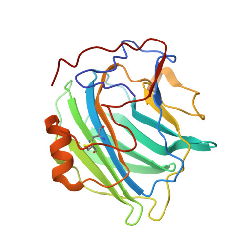
    2W08

  • PubMed Abstract: 

    New therapeutic approaches in Alzheimer's disease are urgently needed. The normal plasma protein, serum amyloid P component (SAP), is always present in cerebrospinal fluid (CSF) and in the pathognomonic lesions of Alzheimer's disease, cerebrovascular and intracerebral Abeta amyloid plaques and neurofibrillary tangles, as a result of its binding to amyloid fibrils and to paired helical filaments, respectively. SAP itself may also be directly neurocytotoxic. Here, in this unique study in Alzheimer's disease of the bis(d-proline) compound, (R)-1-[6-[(R)-2-carboxy-pyrrolidin-1-yl]-6-oxo-hexanoyl]pyrrolidine-2-carboxylic acid (CPHPC), we observed depletion of circulating SAP and also remarkable, almost complete, disappearance of SAP from the CSF. We demonstrate that SAP depletion in vivo is caused by CPHPC cross-linking pairs of SAP molecules in solution to form complexes that are immediately cleared from the plasma. We have also solved the structure of SAP complexed with phosphothreonine, its likely ligand on hyperphosphorylated tau protein. These results support further clinical study of SAP depletion in Alzheimer's disease and potentially other neurodegenerative diseases.


  • Organizational Affiliation
    • Centre for Amyloidosis and Acute Phase Proteins and National Amyloidosis Centre, Division of Medicine (Royal Free Campus), University College London Medical School, London NW3 2PF, United Kingdom.

Macromolecule Content 

  • Total Structure Weight: 118.91 kDa 
  • Atom Count: 9,910 
  • Modeled Residue Count: 1,020 
  • Deposited Residue Count: 1,020 
  • Unique protein chains: 1

Macromolecules

Find similar proteins by:|  3D Structure
Entity ID: 1
MoleculeChains  Sequence LengthOrganismDetailsImage
SERUM AMYLOID P-COMPONENT
A, B, C, D, E
204Homo sapiensMutation(s): 0 
UniProt & NIH Common Fund Data Resources
Find proteins for P02743 (Homo sapiens)
Explore P02743 
Go to UniProtKB:  P02743
PHAROS:  P02743
GTEx:  ENSG00000132703 
Entity Groups
Sequence Clusters30% Identity50% Identity70% Identity90% Identity95% Identity100% Identity
UniProt GroupP02743
Glycosylation
Glycosylation Sites: 1Go to GlyGen: P02743-1
Sequence Annotations
Expand
Reference Sequence

Small Molecules

Ligands 3 Unique
IDChains Name / Formula / InChI Key2D Diagram3D Interactions
NAG

Query on NAG


Download:Ideal Coordinates CCD File
H [auth A],
L [auth B],
P [auth C],
T [auth D],
X [auth E]
2-acetamido-2-deoxy-beta-D-glucopyranose
C8 H15 N O6
OVRNDRQMDRJTHS-FMDGEEDCSA-N
TPO

Query on TPO


Download:Ideal Coordinates CCD File
I [auth A],
M [auth B],
Q [auth C],
U [auth D],
Y [auth E]
PHOSPHOTHREONINE
C4 H10 N O6 P
USRGIUJOYOXOQJ-GBXIJSLDSA-N
CA

Query on CA


Download:Ideal Coordinates CCD File
F [auth A]
G [auth A]
J [auth B]
K [auth B]
N [auth C]
F [auth A],
G [auth A],
J [auth B],
K [auth B],
N [auth C],
O [auth C],
R [auth D],
S [auth D],
V [auth E],
W [auth E]
CALCIUM ION
Ca
BHPQYMZQTOCNFJ-UHFFFAOYSA-N

Experimental Data & Validation

Experimental Data

  • Method: X-RAY DIFFRACTION
  • Resolution: 1.70 Å
  • R-Value Free:  0.176 (Depositor) 
  • R-Value Work:  0.154 (Depositor), 0.151 (DCC) 
  • R-Value Observed: 0.154 (Depositor) 
Space Group: P 1 21 1
Unit Cell:
Length ( Å )Angle ( ˚ )
a = 94.769α = 90
b = 69.435β = 97.05
c = 102.063γ = 90
Software Package:
Software NamePurpose
PHENIXrefinement
MOSFLMdata reduction
SCALAdata scaling
MOLREPphasing

Structure Validation

View Full Validation Report


Ligand Structure Quality Assessment 


Entry History 

Deposition Data

Revision History  (Full details and data files)

  • Version 1.0: 2009-04-14
    Type: Initial release
  • Version 1.1: 2011-05-07
    Changes: Version format compliance
  • Version 1.2: 2011-07-13
    Changes: Version format compliance
  • Version 1.3: 2020-07-29
    Type: Remediation
    Reason: Carbohydrate remediation
    Changes: Advisory, Data collection, Derived calculations, Other, Structure summary
  • Version 1.4: 2023-12-13
    Changes: Data collection, Database references, Refinement description, Structure summary
  • Version 1.5: 2024-10-23
    Changes: Structure summary