2VZV

Substrate Complex of Amycolatopsis orientalis exo-chitosanase CsxA E541A with chitosan


Experimental Data Snapshot

  • Method: X-RAY DIFFRACTION
  • Resolution: 2.70 Å
  • R-Value Free: 0.255 
  • R-Value Work: 0.182 
  • R-Value Observed: 0.186 

wwPDB Validation   3D Report Full Report


This is version 2.0 of the entry. See complete history


Literature

The Structural Basis of Substrate Recognition in an Exo-Beta-D-Glucosaminidase Involved in Chitosan Hydrolysis.

Lammerts Van Bueren, A.Ghinet, M.G.Gregg, K.Fleury, A.Brzezinski, R.Boraston, A.B.

(2009) J Mol Biol 385: 131

  • DOI: https://doi.org/10.1016/j.jmb.2008.10.031
  • Primary Citation of Related Structures:  
    2VZO, 2VZS, 2VZT, 2VZU, 2VZV

  • PubMed Abstract: 

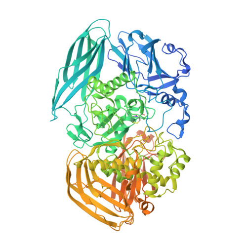
    Family 2 of the glycoside hydrolase classification is one of the largest families. Structurally characterized members of this family include enzymes with beta-galactosidase activity (Escherichia coli LacZ), beta-glucuronidase activity (Homo sapiens GusB), and beta-mannosidase activity (Bacteroides thetaiotaomicron BtMan2A). Here, we describe the structure of a family 2 glycoside hydrolase, CsxA, from Amycolatopsis orientalis that has exo-beta-D-glucosaminidase (exo-chitosanase) activity. Analysis of a product complex (1.85 A resolution) reveals a unique negatively charged pocket that specifically accommodates the nitrogen of nonreducing end glucosamine residues, allowing this enzyme to discriminate between glucose and glucosamine. This also provides structural evidence for the role of E541 as the catalytic nucleophile and D469 as the catalytic acid/base. The structures of an E541A mutant in complex with a natural beta-1,4-D-glucosamine tetrasaccharide substrate and both E541A and D469A mutants in complex with a pNP-beta-D-glucosaminide synthetic substrate provide insight into interactions in the +1 subsite of this enzyme. Overall, a comparison with the active sites of other GH2 enzymes highlights the unique architecture of the CsxA active site, which imparts specificity for its cationic substrate.


  • Organizational Affiliation

    Biochemistry and Microbiology, University of Victoria, BC, Canada.


Macromolecules
Find similar proteins by:  (by identity cutoff)  |  3D Structure
Entity ID: 1
MoleculeChains Sequence LengthOrganismDetailsImage
EXO-BETA-D-GLUCOSAMINIDASE
A, B
1,032Amycolatopsis orientalisMutation(s): 1 
UniProt
Find proteins for Q56F26 (Amycolatopsis orientalis)
Explore Q56F26 
Go to UniProtKB:  Q56F26
Entity Groups  
Sequence Clusters30% Identity50% Identity70% Identity90% Identity95% Identity100% Identity
UniProt GroupQ56F26
Sequence Annotations
Expand
  • Reference Sequence
Oligosaccharides

Help

Entity ID: 2
MoleculeChains Length2D Diagram Glycosylation3D Interactions
2-amino-2-deoxy-beta-D-glucopyranose-(1-4)-2-amino-2-deoxy-beta-D-glucopyranose
C, D
2N/A
Glycosylation Resources
GlyTouCan:  G61071JD
GlyCosmos:  G61071JD
GlyGen:  G61071JD
Experimental Data & Validation

Experimental Data

  • Method: X-RAY DIFFRACTION
  • Resolution: 2.70 Å
  • R-Value Free: 0.255 
  • R-Value Work: 0.182 
  • R-Value Observed: 0.186 
  • Space Group: P 21 21 21
Unit Cell:
Length ( Å )Angle ( ˚ )
a = 86.86α = 90
b = 121.41β = 90
c = 184.71γ = 90
Software Package:
Software NamePurpose
REFMACrefinement

Structure Validation

View Full Validation Report


Entry History 

Deposition Data

Revision History  (Full details and data files)

  • Version 1.0: 2008-10-14
    Type: Initial release
  • Version 1.1: 2011-08-17
    Changes: Atomic model, Derived calculations, Non-polymer description, Other, Refinement description, Structure summary, Version format compliance
  • Version 2.0: 2020-07-29
    Type: Remediation
    Reason: Carbohydrate remediation
    Changes: Advisory, Atomic model, Data collection, Derived calculations, Other, Structure summary