Development and Validation of a Cytochrome C Coupled Assay for Pteridine Reductase 1 and Dihydrofolate Reductase.
Shanks, E.J., Ong, H.B., Robinson, D.A., Thompson, S., Sienkiewicz, N., Fairlamb, A.H., Frearson, J.A.(2010) Anal Biochem 396: 194
- PubMed: 19748480 Search on PubMedSearch on PubMed Central
- DOI: https://doi.org/10.1016/j.ab.2009.09.003
- Primary Citation Related Structures:
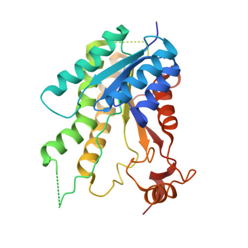
2VZ0 - PubMed Abstract:
Activity of the pterin- and folate-salvaging enzymes pteridine reductase 1 (PTR1) and dihydrofolate reductase-thymidylate synthetase (DHFR-TS) is commonly measured as a decrease in absorbance at 340 nm, corresponding to oxidation of nicotinamide adenine dinucleotide phosphate (NADPH). Although this assay has been adequate to study the biology of these enzymes, it is not amenable to support any degree of routine inhibitor assessment because its restricted linearity is incompatible with enhanced throughput microtiter plate screening. In this article, we report the development and validation of a nonenzymatically coupled screening assay in which the product of the enzymatic reaction reduces cytochrome c, causing an increase in absorbance at 550 nm. We demonstrate this assay to be robust and accurate, and we describe its utility in supporting a structure-based design, small-molecule inhibitor campaign against Trypanosoma brucei PTR1 and DHFR-TS.
- Division of Biological Chemistry and Drug Discovery, College of Life Sciences, University of Dundee, Dundee DD15EH, UK.
Organizational Affiliation: