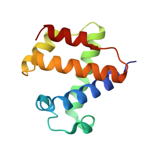
2VYY

Mutant Ala55Trp of Cerebratuls lacteus mini-hemoglobin


Experimental Data Snapshot

  • Method: X-RAY DIFFRACTION
  • Resolution: 1.60 Å
  • R-Value Free: 0.213 
  • R-Value Work: 0.167 
  • R-Value Observed: 0.171 

Starting Model: experimental
View more details

wwPDB Validation   3D Report Full Report


Ligand Structure Quality Assessment 


This is version 1.3 of the entry. See complete history


Literature

The Apolar Channel in Cerebratulus Lacteus Hemoglobin is the Route for O2 Entry and Exit.

Salter, M.D.Nienhaus, K.Nienhaus, G.U.Dewilde, S.Moens, L.Pesce, A.Nardini, M.Bolognesi, M.Olson, J.S.

(2008) J Biol Chem 283: 35689

  • DOI: https://doi.org/10.1074/jbc.M805727200
  • Primary Citation of Related Structures:  
    2VYY, 2VYZ

  • PubMed Abstract: 

    The major pathway for O2 binding to mammalian myoglobins (Mb) and hemoglobins (Hb) involves transient upward movement of the distal histidine (His-64(E7)), allowing ligand capture in the distal pocket. The mini-globin from Cerebratulus lacteus (CerHb) appears to have an alternative pathway between the E and H helices that is made accessible by loss of the N-terminal A helix. To test this pathway, we examined the effects of changing the size of the E7 gate and closing the end of the apolar channel in CerHb by site-directed mutagenesis. Increasing the size of Gln-44(E7) from Ala to Trp causes variation of association (k'O2) and dissociation (kO2) rate coefficients, but the changes are not systematic. More significantly, the fractions (Fgem approximately 0.05-0.19) and rates (kgem approximately 50-100 micros(-1)) of geminate CO recombination in the Gln-44(E7) mutants are all similar. In contrast, blocking the entrance to the apolar channel by increasing the size of Ala-55(E18) to Phe and Trp causes the following: 1) both k'O2 and kO2 to decrease roughly 4-fold; 2) Fgem for CO to increase from approximately 0.05 to 0.45; and 3) kgem to decrease from approximately 80 to approximately 9 micros(-1), as ligands become trapped in the channel. Crystal structures and low temperature Fourier-transform infrared spectra of Phe-55 and Trp-55 CerHb confirm that the aromatic side chains block the channel entrance, with little effect on the distal pocket. These results provide unambiguous experimental proof that diatomic ligands can enter and exit a globin through an interior channel in preference to the more direct E7 pathway.


  • Organizational Affiliation

    Department of Biochemistry and Cell Biology, Rice University, Houston, Texas 77005-1892, USA.


Macromolecules
Find similar proteins by:  (by identity cutoff)  |  3D Structure
Entity ID: 1
MoleculeChains Sequence LengthOrganismDetailsImage
NEURAL HEMOGLOBIN110Cerebratulus lacteusMutation(s): 1 
UniProt
Find proteins for O76242 (Cerebratulus lacteus)
Explore O76242 
Go to UniProtKB:  O76242
Entity Groups  
Sequence Clusters30% Identity50% Identity70% Identity90% Identity95% Identity100% Identity
UniProt GroupO76242
Sequence Annotations
Expand
  • Reference Sequence
Small Molecules
Ligands 4 Unique
IDChains Name / Formula / InChI Key2D Diagram3D Interactions
HEM
Query on HEM

Download Ideal Coordinates CCD File 
B [auth A]PROTOPORPHYRIN IX CONTAINING FE
C34 H32 Fe N4 O4
KABFMIBPWCXCRK-RGGAHWMASA-L
SO4
Query on SO4

Download Ideal Coordinates CCD File 
D [auth A],
E [auth A]
SULFATE ION
O4 S
QAOWNCQODCNURD-UHFFFAOYSA-L
GOL
Query on GOL

Download Ideal Coordinates CCD File 
F [auth A],
G [auth A]
GLYCEROL
C3 H8 O3
PEDCQBHIVMGVHV-UHFFFAOYSA-N
OXY
Query on OXY

Download Ideal Coordinates CCD File 
C [auth A]OXYGEN MOLECULE
O2
MYMOFIZGZYHOMD-UHFFFAOYSA-N
Experimental Data & Validation

Experimental Data

  • Method: X-RAY DIFFRACTION
  • Resolution: 1.60 Å
  • R-Value Free: 0.213 
  • R-Value Work: 0.167 
  • R-Value Observed: 0.171 
  • Space Group: P 21 21 21
Unit Cell:
Length ( Å )Angle ( ˚ )
a = 42.79α = 90
b = 43.323β = 90
c = 60.036γ = 90
Software Package:
Software NamePurpose
REFMACrefinement
MOSFLMdata reduction
SCALAdata scaling
MOLREPphasing

Structure Validation

View Full Validation Report


Ligand Structure Quality Assessment 


Entry History 

Deposition Data

Revision History  (Full details and data files)

  • Version 1.0: 2008-10-07
    Type: Initial release
  • Version 1.1: 2013-04-17
    Changes: Database references, Derived calculations, Non-polymer description, Other, Refinement description, Structure summary, Version format compliance
  • Version 1.2: 2019-03-06
    Changes: Advisory, Data collection, Experimental preparation, Other
  • Version 1.3: 2023-12-13
    Changes: Advisory, Data collection, Database references, Derived calculations, Other, Refinement description