2VXS

Structure of IL-17A in complex with a potent, fully human neutralising antibody


Experimental Data Snapshot

  • Method: X-RAY DIFFRACTION
  • Resolution: 2.63 Å
  • R-Value Free: 0.264 
  • R-Value Work: 0.213 
  • R-Value Observed: 0.215 

Starting Model: experimental
View more details

wwPDB Validation   3D Report Full Report


This is version 1.3 of the entry. See complete history


Literature

Structure of Il-17A in Complex with a Potent, Fully Human Neutralising Antibody.

Gerhardt, S.Abbott, W.M.Hargreaves, D.Pauptit, R.A.Davies, R.A.Needham, M.R.Langham, C.Barker, W.Aziz, A.Snow, M.J.Dawson, S.Welsh, F.Wilkinson, T.Vaugan, T.Beste, G.Bishop, S.Popovic, B.Rees, G.Sleeman, M.Tuske, S.J.Coales, S.J.Hamuro, Y.Russell, C.

(2009) J Mol Biol 394: 905

  • DOI: https://doi.org/10.1016/j.jmb.2009.10.008
  • Primary Citation of Related Structures:  
    2VXS

  • PubMed Abstract: 

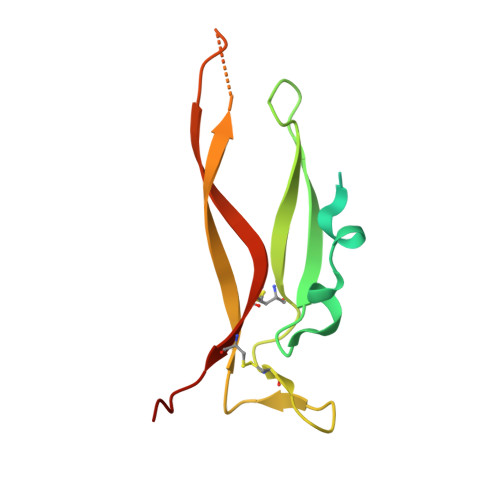
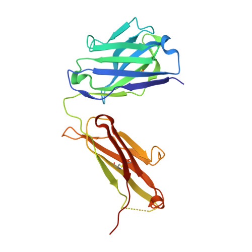
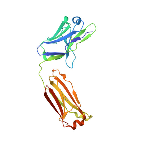
    IL-17A is a pro-inflammatory cytokine produced by the newly identified Th17 subset of T-cells. We have isolated a human monoclonal antibody to IL-17A (CAT-2200) that can potently neutralize the effects of recombinant and native human IL-17A. We determined the crystal structure of IL-17A in complex with the CAT-2200 Fab at 2.6 A resolution in order to provide a definitive characterization of the epitope and paratope regions. Approximately a third of the IL-17A dimer is disordered in this crystal structure. The disorder occurs in both independent copies of the complex in the asymmetric unit and does not appear to be influenced by crystal packing. The complex contains one IL-17A dimer sandwiched between two CAT-2200 Fab fragments. The IL-17A is a disulfide-linked homodimer that is similar in structure to IL-17F, adopting a cystine-knot fold. The structure is not inconsistent with the previous prediction of a receptor binding cavity on IL-17 family members. The epitope recognized by CAT-2200 is shown to involve 12 amino acid residues from the quaternary structure of IL-17A, with each Fab contacting both monomers in the dimer. All complementarity-determining regions (CDRs) in the Fab contribute to a total of 16 amino acid residues in the antibody paratope. In vitro affinity optimization was used to generate CAT-2200 from a parental lead antibody using random mutagenesis of CDR3 loops. This resulted in seven amino acid changes (three in VL-CDR3 and four in VH-CDR3) and gave an approximate 30-fold increase in potency in a cell-based neutralization assay. Two of the seven amino acids form part of the CAT-2200 paratope. The observed interaction site between CAT-2200 and IL-17A is consistent with data from hydrogen/deuterium exchange mass spectrometry and mutagenesis approaches.


  • Organizational Affiliation

    AstraZeneca, Alderley Park, Macclesfield, Cheshire SK10 4TG, UK.


Macromolecules
Find similar proteins by:  (by identity cutoff)  |  3D Structure
Entity ID: 1
MoleculeChains Sequence LengthOrganismDetailsImage
INTERLEUKIN-17A
A, B, C, D
137Homo sapiensMutation(s): 0 
UniProt & NIH Common Fund Data Resources
Find proteins for Q16552 (Homo sapiens)
Explore Q16552 
Go to UniProtKB:  Q16552
PHAROS:  Q16552
GTEx:  ENSG00000112115 
Entity Groups  
Sequence Clusters30% Identity50% Identity70% Identity90% Identity95% Identity100% Identity
UniProt GroupQ16552
Sequence Annotations
Expand
  • Reference Sequence
Find similar proteins by:  (by identity cutoff)  |  3D Structure
Entity ID: 2
MoleculeChains Sequence LengthOrganismDetailsImage
FAB FRAGMENTE [auth H],
F [auth I],
G [auth J],
H [auth K]
225Homo sapiensMutation(s): 0 
Entity Groups  
Sequence Clusters30% Identity50% Identity70% Identity90% Identity95% Identity100% Identity
Sequence Annotations
Expand
  • Reference Sequence
Find similar proteins by:  (by identity cutoff)  |  3D Structure
Entity ID: 3
MoleculeChains Sequence LengthOrganismDetailsImage
FAB FRAGMENTI [auth L],
J [auth M],
K [auth N],
L [auth O]
216Homo sapiensMutation(s): 0 
Entity Groups  
Sequence Clusters30% Identity50% Identity70% Identity90% Identity95% Identity100% Identity
Sequence Annotations
Expand
  • Reference Sequence
Small Molecules
Ligands 1 Unique
IDChains Name / Formula / InChI Key2D Diagram3D Interactions
SO4
Query on SO4

Download Ideal Coordinates CCD File 
M [auth A]
N [auth B]
O [auth B]
P [auth C]
Q [auth D]
M [auth A],
N [auth B],
O [auth B],
P [auth C],
Q [auth D],
R [auth I],
S [auth I],
T [auth L],
U [auth L],
V [auth M],
W [auth N],
X [auth O]
SULFATE ION
O4 S
QAOWNCQODCNURD-UHFFFAOYSA-L
Experimental Data & Validation

Experimental Data

  • Method: X-RAY DIFFRACTION
  • Resolution: 2.63 Å
  • R-Value Free: 0.264 
  • R-Value Work: 0.213 
  • R-Value Observed: 0.215 
  • Space Group: P 1 21 1
Unit Cell:
Length ( Å )Angle ( ˚ )
a = 98.473α = 90
b = 66.708β = 91.65
c = 203.79γ = 90
Software Package:
Software NamePurpose
REFMACrefinement
MOSFLMdata reduction
SCALAdata scaling
MOLREPphasing

Structure Validation

View Full Validation Report


Entry History 

Revision History  (Full details and data files)

  • Version 1.0: 2009-07-14
    Type: Initial release
  • Version 1.1: 2011-07-13
    Changes: Advisory, Version format compliance
  • Version 1.2: 2023-12-13
    Changes: Data collection, Database references, Derived calculations, Other, Refinement description
  • Version 1.3: 2024-10-23
    Changes: Structure summary