2VXH | pdb_00002vxh

The crystal structure of chlorite dismutase: a detox enzyme producing molecular oxygen


Experimental Data Snapshot

  • Method: X-RAY DIFFRACTION
  • Resolution: 2.10 Å
  • R-Value Free: 
    0.255 (Depositor), 0.253 (DCC) 
  • R-Value Work: 
    0.219 (Depositor), 0.218 (DCC) 
  • R-Value Observed: 
    0.221 (Depositor) 

wwPDB Validation 3D Report Full Report

Validation slider image for 2VXH

Ligand Structure Quality Assessment 


This is version 1.3 of the entry. See complete history

Literature

Crystal Structure of Chlorite Dismutase, a Detoxifying Enzyme Producing Molecular Oxygen

De Geus, D.C.Thomassen, E.A.J.Hagedoorn, P.L.Pannu, N.S.Van Duijn, E.Abrahams, J.P.

(2009) J Mol Biology 387: 192

  • DOI: https://doi.org/10.1016/j.jmb.2009.01.036
  • Primary Citation Related Structures: 
    2VXH

  • PubMed Abstract: 

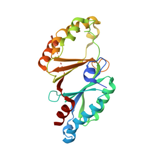
    Chlorite dismutase (Cld) is a key enzyme of perchlorate and chlorate respiration. This heme-based protein reduces the toxic compound chlorite into the innocuous chloride anion in a very efficient way while producing molecular oxygen. A sequence comparison between Cld homologues shows a highly conserved family. The crystal structure of Azospira oryzae strain GR-1 Cld is reported to 2.1 A resolution. The structure reveals a hexameric organization of the Cld, while each monomer exhibits a ferredoxin-like fold. The six subunits are organized in a ring structure with a maximal diameter of 9 nm and an inner diameter of 2 nm. The heme active-site pocket is solvent accessible both from the inside and the outside of the ring. Moreover, a second anion binding site that could accommodate the assumed reaction intermediate ClO(-) for further transformation has been identified near the active site. The environment of the heme cofactor was investigated with electron paramagnetic resonance spectroscopy. Apart from the high-spin ferric signal of the five-coordinate resting-state enzyme, two low-spin signals were found corresponding to six-coordinate species. The current crystal structure confirms and complements a recently proposed catalytic mechanism that proceeds via a ferryl species and a ClO(-) anion. Our structural data exclude cooperativity between the iron centers.


  • Organizational Affiliation
    • Department of Biophysical Structural Chemistry, Leiden Institute of Chemistry, Leiden University, Leiden, The Netherlands. d.de.geus@chem.leidenuniv.nl

Macromolecule Content 

  • Total Structure Weight: 175.3 kDa 
  • Atom Count: 11,790 
  • Modeled Residue Count: 1,375 
  • Deposited Residue Count: 1,506 
  • Unique protein chains: 1

Macromolecules

Find similar proteins by:|  3D Structure
Entity ID: 1
MoleculeChains  Sequence LengthOrganismDetailsImage
CHLORITE DISMUTASE
A, B, C, D, E
A, B, C, D, E, F
251Azospira oryzaeMutation(s): 0 
EC: 1.13.11.49
UniProt
Find proteins for E2DI02 (Azospira oryzae)
Explore E2DI02 
Go to UniProtKB:  E2DI02
Entity Groups
Sequence Clusters30% Identity50% Identity70% Identity90% Identity95% Identity100% Identity
UniProt GroupE2DI02
Sequence Annotations
Expand
Reference Sequence

Small Molecules

Ligands 3 Unique
IDChains Name / Formula / InChI Key2D Diagram3D Interactions
HEM

Query on HEM


Download:Ideal Coordinates CCD File
G [auth A]
J [auth B]
M [auth C]
P [auth D]
R [auth E]
G [auth A],
J [auth B],
M [auth C],
P [auth D],
R [auth E],
U [auth F]
PROTOPORPHYRIN IX CONTAINING FE
C34 H32 Fe N4 O4
KABFMIBPWCXCRK-RGGAHWMASA-L
CO3

Query on CO3


Download:Ideal Coordinates CCD File
I [auth A],
L [auth B],
O [auth C],
T [auth E],
W [auth F]
CARBONATE ION
C O3
BVKZGUZCCUSVTD-UHFFFAOYSA-L
SCN

Query on SCN


Download:Ideal Coordinates CCD File
H [auth A]
K [auth B]
N [auth C]
Q [auth D]
S [auth E]
H [auth A],
K [auth B],
N [auth C],
Q [auth D],
S [auth E],
V [auth F]
THIOCYANATE ION
C N S
ZMZDMBWJUHKJPS-UHFFFAOYSA-M

Experimental Data & Validation

Experimental Data

  • Method: X-RAY DIFFRACTION
  • Resolution: 2.10 Å
  • R-Value Free:  0.255 (Depositor), 0.253 (DCC) 
  • R-Value Work:  0.219 (Depositor), 0.218 (DCC) 
  • R-Value Observed: 0.221 (Depositor) 
Space Group: P 21 21 2
Unit Cell:
Length ( Å )Angle ( ˚ )
a = 164.458α = 90
b = 169.335β = 90
c = 60.792γ = 90
Software Package:
Software NamePurpose
REFMACrefinement
MOSFLMdata reduction
SCALAdata scaling
CRANKphasing

Structure Validation

View Full Validation Report


Ligand Structure Quality Assessment 


Entry History 

Deposition Data

Revision History  (Full details and data files)

  • Version 1.0: 2009-03-03
    Type: Initial release
  • Version 1.1: 2011-05-07
    Changes: Version format compliance
  • Version 1.2: 2011-07-13
    Changes: Version format compliance
  • Version 1.3: 2024-05-08
    Changes: Data collection, Database references, Derived calculations, Other, Refinement description