2VWQ | pdb_00002vwq

Haloferax mediterranei glucose dehydrogenase in complex with NADP and Zn.


Experimental Data Snapshot

  • Method: X-RAY DIFFRACTION
  • Resolution: 2.10 Å
  • R-Value Free: 
    0.175 (Depositor), 0.188 (DCC) 
  • R-Value Work: 
    0.143 (Depositor), 0.155 (DCC) 
  • R-Value Observed: 
    0.145 (Depositor) 

Starting Model: experimental
View more details

wwPDB Validation 3D Report Full Report

Validation slider image for 2VWQ

Ligand Structure Quality Assessment 


This is version 1.4 of the entry. See complete history

Literature

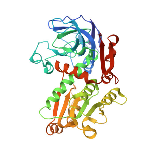
Active site dynamics in the zinc-dependent medium chain alcohol dehydrogenase superfamily.

Baker, P.J.Britton, K.L.Fisher, M.Esclapez, J.Pire, C.Bonete, M.J.Ferrer, J.Rice, D.W.

(2009) Proc Natl Acad Sci U S A 106: 779-784

  • DOI: https://doi.org/10.1073/pnas.0807529106
  • Primary Citation Related Structures: 
    2VWG, 2VWH, 2VWP, 2VWQ

  • PubMed Abstract: 

    Despite being the subject of intensive investigations, many aspects of the mechanism of the zinc-dependent medium chain alcohol dehydrogenase (MDR) superfamily remain contentious. We have determined the high-resolution structures of a series of binary and ternary complexes of glucose dehydrogenase, an MDR enzyme from Haloferax mediterranei. In stark contrast to the textbook MDR mechanism in which the zinc ion is proposed to remain stationary and attached to a common set of protein ligands, analysis of these structures reveals that in each complex, there are dramatic differences in the nature of the zinc ligation. These changes arise as a direct consequence of linked movements of the zinc ion, a zinc-bound bound water molecule, and the substrate during progression through the reaction. These results provide evidence for the molecular basis of proton traffic during catalysis, a structural explanation for pentacoordinate zinc ion intermediates, a unifying view for the observed patterns of metal ligation in the MDR family, and highlight the importance of dynamic fluctuations at the metal center in changing the electrostatic potential in the active site, thereby influencing the proton traffic and hydride transfer events.


  • Organizational Affiliation
    • The Krebs Institute, Department of Molecular Biology and Biotechnology, University of Sheffield, Western Bank, Sheffield S10 2TN, United Kingdom. P.Baker@sheffield.ac.uk

Macromolecule Content 

  • Total Structure Weight: 40.09 kDa 
  • Atom Count: 2,932 
  • Modeled Residue Count: 357 
  • Deposited Residue Count: 357 
  • Unique protein chains: 1

Macromolecules

Find similar proteins by:|  3D Structure
Entity ID: 1
MoleculeChains  Sequence LengthOrganismDetailsImage
GLUCOSE DEHYDROGENASE357Haloferax mediterraneiMutation(s): 0 
EC: 1.1.1.47
UniProt
Find proteins for Q977U7 (Haloferax mediterranei (strain ATCC 33500 / DSM 1411 / JCM 8866 / NBRC 14739 / NCIMB 2177 / R-4))
Explore Q977U7 
Go to UniProtKB:  Q977U7
Entity Groups
Sequence Clusters30% Identity50% Identity70% Identity90% Identity95% Identity100% Identity
UniProt GroupQ977U7
Sequence Annotations
Expand
Reference Sequence

Small Molecules

Ligands 2 Unique
IDChains Name / Formula / InChI Key2D Diagram3D Interactions
NAP

Query on NAP


Download:Ideal Coordinates CCD File
B [auth A]NADP NICOTINAMIDE-ADENINE-DINUCLEOTIDE PHOSPHATE
C21 H28 N7 O17 P3
XJLXINKUBYWONI-NNYOXOHSSA-N
ZN

Query on ZN


Download:Ideal Coordinates CCD File
C [auth A]ZINC ION
Zn
PTFCDOFLOPIGGS-UHFFFAOYSA-N

Experimental Data & Validation

Experimental Data

  • Method: X-RAY DIFFRACTION
  • Resolution: 2.10 Å
  • R-Value Free:  0.175 (Depositor), 0.188 (DCC) 
  • R-Value Work:  0.143 (Depositor), 0.155 (DCC) 
  • R-Value Observed: 0.145 (Depositor) 
Space Group: I 2 2 2
Unit Cell:
Length ( Å )Angle ( ˚ )
a = 61.884α = 90
b = 111.113β = 90
c = 152.227γ = 90
Software Package:
Software NamePurpose
REFMACrefinement
DENZOdata reduction
SCALEPACKdata scaling
AMoREphasing

Structure Validation

View Full Validation Report


Ligand Structure Quality Assessment 


Entry History 

Deposition Data

Revision History  (Full details and data files)

  • Version 1.0: 2009-01-13
    Type: Initial release
  • Version 1.1: 2011-05-08
    Changes: Version format compliance
  • Version 1.2: 2011-07-13
    Changes: Version format compliance
  • Version 1.3: 2017-08-09
    Changes: Database references
  • Version 1.4: 2023-12-13
    Changes: Data collection, Database references, Derived calculations, Other, Refinement description