2VRY

Mouse Neuroglobin with heme iron in the reduced ferrous state


Experimental Data Snapshot

  • Method: X-RAY DIFFRACTION
  • Resolution: 1.87 Å
  • R-Value Free: 0.217 
  • R-Value Work: 0.185 
  • R-Value Observed: 0.187 

wwPDB Validation   3D Report Full Report


Ligand Structure Quality Assessment 


This is version 1.4 of the entry. See complete history


Literature

An X-Ray Diffraction and X-Ray Absorption Spectroscopy Joint Study of Neuroglobin.

Arcovito, A.Moschetti, T.D'Angelo, P.Mancini, G.Vallone, B.Brunori, M.Della Longa, S.

(2008) Arch Biochem Biophys 475: 7

  • DOI: https://doi.org/10.1016/j.abb.2008.03.026
  • Primary Citation of Related Structures:  
    2VRY

  • PubMed Abstract: 

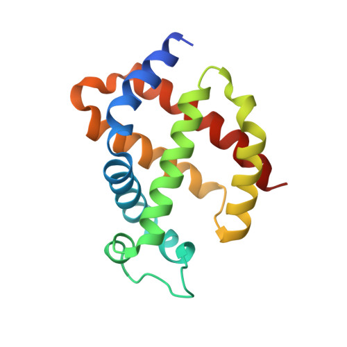
    Neuroglobin (Ngb) is a member of the globin family expressed in the vertebrate brain, involved in neuroprotection. A combined approach of X-ray diffraction (XRD) on single crystal and X-ray absorption spectroscopy (XAS) in solution, allows to determine the oxidation state and the structure of the Fe-heme both in the bis-histidine and the CO-bound (NgbCO) states. The overall data demonstrate that under X-ray the iron is photoreduced fairly rapidly, and that the previously reported X-ray structure of ferric Ngb [B. Vallone, K. Nienhaus, M. Brunori, G.U. Nienhaus, Proteins 56 (2004) 85-92] very likely refers to a photoreduced species indistinguishable from the dithionite reduced protein. Results from the XAS analysis of NgbCO in solution are in good agreement with XRD data on the crystal. However prolonged X-ray exposure at 15K determines CO release. This preliminary result paves the way to experiments aimed at the characterization of pentacoordinate ferrous Ngb, the only species competent in binding external ligands such as O2, CO or NO.


  • Organizational Affiliation

    Istituto di Biochimica e Biochimica Clinica, Università Cattolica del Sacro Cuore, Largo F. Vito 1, 00167 Rome, Italy.


Macromolecules
Find similar proteins by:  (by identity cutoff)  |  3D Structure
Entity ID: 1
MoleculeChains Sequence LengthOrganismDetailsImage
NEUROGLOBIN154Mus musculusMutation(s): 2 
UniProt
Find proteins for Q9ER97 (Mus musculus)
Explore Q9ER97 
Go to UniProtKB:  Q9ER97
Entity Groups  
Sequence Clusters30% Identity50% Identity70% Identity90% Identity95% Identity100% Identity
UniProt GroupQ9ER97
Sequence Annotations
Expand
  • Reference Sequence
Experimental Data & Validation

Experimental Data

  • Method: X-RAY DIFFRACTION
  • Resolution: 1.87 Å
  • R-Value Free: 0.217 
  • R-Value Work: 0.185 
  • R-Value Observed: 0.187 
  • Space Group: H 3 2
Unit Cell:
Length ( Å )Angle ( ˚ )
a = 87.92α = 90
b = 87.92β = 90
c = 113.646γ = 120
Software Package:
Software NamePurpose
REFMACrefinement
HKL-2000data reduction
SCALEPACKdata scaling
REFMACphasing

Structure Validation

View Full Validation Report


Ligand Structure Quality Assessment 


Entry History 

Deposition Data

Revision History  (Full details and data files)

  • Version 1.0: 2008-05-06
    Type: Initial release
  • Version 1.1: 2011-07-13
    Changes: Advisory, Version format compliance
  • Version 1.2: 2014-10-22
    Changes: Database references
  • Version 1.3: 2019-03-06
    Changes: Data collection, Experimental preparation, Other
  • Version 1.4: 2023-12-13
    Changes: Data collection, Database references, Derived calculations, Other, Refinement description