2VRT

Crystal Structure of E. coli RNase E possessing M1 RNA fragments - Catalytic Domain


Experimental Data Snapshot

  • Method: X-RAY DIFFRACTION
  • Resolution: 3.50 Å
  • R-Value Free: 0.351 
  • R-Value Work: 0.317 
  • R-Value Observed: 0.321 

wwPDB Validation   3D Report Full Report


This is version 1.3 of the entry. See complete history


Literature

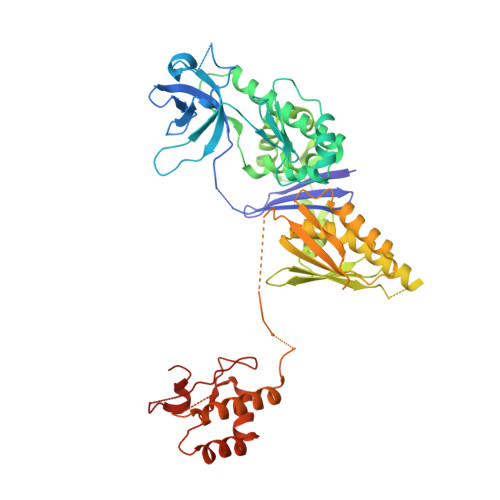
The Crystal Structure of the Escherichia Coli Rnase E Apoprotein and a Mechanism for RNA Degradation.

Koslover, D.J.Callaghan, A.J.Marcaida, M.J.Garman, E.F.Martick, M.Scott, W.G.Luisi, B.F.

(2008) Structure 16: 1238

  • DOI: https://doi.org/10.1016/j.str.2008.04.017
  • Primary Citation of Related Structures:  
    2VMK, 2VRT

  • PubMed Abstract: 

    RNase E is an essential bacterial endoribonuclease involved in the turnover of messenger RNA and the maturation of structured RNA precursors in Escherichia coli. Here, we present the crystal structure of the E. coli RNase E catalytic domain in the apo-state at 3.3 A. This structure indicates that, upon catalytic activation, RNase E undergoes a marked conformational change characterized by the coupled movement of two RNA-binding domains to organize the active site. The structural data suggest a mechanism of RNA recognition and cleavage that explains the enzyme's preference for substrates possessing a 5'-monophosphate and accounts for the protective effect of a triphosphate cap for most transcripts. Internal flexibility within the quaternary structure is also observed, a finding that has implications for recognition of structured RNA substrates and for the mechanism of internal entry for a subset of substrates that are cleaved without 5'-end requirements.


  • Organizational Affiliation

    Department of Biochemistry, University of Cambridge, Tennis Court Road, Cambridge CB2 1GA, United Kingdom.


Macromolecules

Find similar proteins by:  (by identity cutoff)  |  3D Structure
Entity ID: 1
MoleculeChains Sequence LengthOrganismDetailsImage
RIBONUCLEASE E
A, B, C, D
509Escherichia coliMutation(s): 0 
EC: 3.1.4
UniProt
Find proteins for P21513 (Escherichia coli (strain K12))
Explore P21513 
Go to UniProtKB:  P21513
Entity Groups  
Sequence Clusters30% Identity50% Identity70% Identity90% Identity95% Identity100% Identity
UniProt GroupP21513
Sequence Annotations
Expand
  • Reference Sequence

Find similar nucleic acids by:  Sequence   |   3D Structure  

Entity ID: 2
MoleculeChains LengthOrganismImage
5'-R(*UP*UP)-3'
E, F
2Escherichia coli
Sequence Annotations
Expand
  • Reference Sequence

Find similar nucleic acids by:  Sequence   |   3D Structure  

Entity ID: 3
MoleculeChains LengthOrganismImage
5'-R(*UP*UP*GP)-3'
G, H
3Escherichia coli
Sequence Annotations
Expand
  • Reference Sequence
Experimental Data & Validation

Experimental Data

  • Method: X-RAY DIFFRACTION
  • Resolution: 3.50 Å
  • R-Value Free: 0.351 
  • R-Value Work: 0.317 
  • R-Value Observed: 0.321 
  • Space Group: C 1 2 1
Unit Cell:
Length ( Å )Angle ( ˚ )
a = 93.894α = 90
b = 121.252β = 99.47
c = 242.228γ = 90
Software Package:
Software NamePurpose
REFMACrefinement
MOSFLMdata reduction
SCALAdata scaling
PHASERphasing
MOLREPphasing
REFMACphasing

Structure Validation

View Full Validation Report


Entry History 

Deposition Data

Revision History  (Full details and data files)

  • Version 1.0: 2008-07-22
    Type: Initial release
  • Version 1.1: 2011-05-08
    Changes: Version format compliance
  • Version 1.2: 2011-07-13
    Changes: Version format compliance
  • Version 1.3: 2023-12-13
    Changes: Data collection, Database references, Derived calculations, Other, Refinement description