2VQG

Crystal structure of PorB from Corynebacterium glutamicum (crystal form I)


Experimental Data Snapshot

  • Method: X-RAY DIFFRACTION
  • Resolution: 1.82 Å
  • R-Value Free: 0.215 
  • R-Value Work: 0.195 
  • R-Value Observed: 0.196 

wwPDB Validation   3D Report Full Report


Ligand Structure Quality Assessment 


This is version 1.1 of the entry. See complete history


Literature

A Putative Alpha-Helical Porin from Corynebacterium Glutamicum.

Ziegler, K.Benz, R.Schulz, G.E.

(2008) J Mol Biol 379: 482

  • DOI: https://doi.org/10.1016/j.jmb.2008.04.017
  • Primary Citation of Related Structures:  
    2VQG, 2VQH, 2VQK, 2VQL

  • PubMed Abstract: 

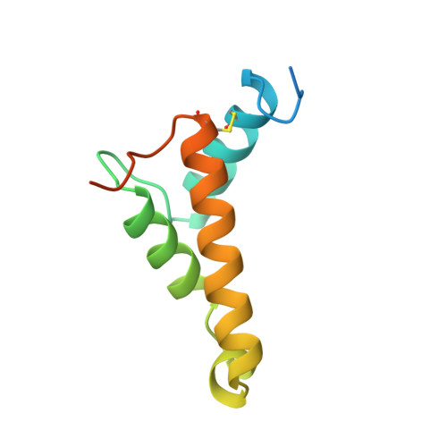
    The cell wall of Corynebacterium glutamicum contains a mycolic acid layer, which is a protective nonpolar barrier similar to the outer membrane of Gram-negative bacteria. The exchange of material across this barrier requires porins. Porin B (PorB) is one of them. Recombinant PorB has been produced in Escherichia coli, purified, crystallized and analyzed by X-ray diffraction, yielding 16 independent molecular structures in four different crystal forms at resolutions up to 1.8 A. All 16 molecules have the same globular core, which consists of 70 residues forming four alpha-helices tied together by a disulfide bridge. The 16 structures vary greatly with respect to the 29 residues in the N- and C-terminal extensions. Since corynebacteria belong to the group of mycolata that includes some prominent human pathogens, the observed structure may be of medical relevance. Due to the clearly established solid structure of the core, the native porin has to be oligomeric, and the reported structure is one of the subunits. An alpha-helical porin in a bacterial outer envelope is surprising because all presently known structures of such porins consist of beta-barrels. Since none of the four crystal packing arrangements was compatible with an oligomeric membrane channel, we constructed a model of such an oligomer that was consistent with all available data of native PorB. The proposed model is based on the required polar interior and nonpolar exterior of the porin, on a recurring crystal packing contact around a 2-fold axis, on the assumption of a simple C(n) symmetry (a symmetric arrangement around an n-fold axis), on the experimentally established electric conductivity and anion selectivity and on the generally observed shape of porin channels.


  • Organizational Affiliation

    Institut für Organische Chemie und Biochemie, Albert-Ludwigs-Universität, Albertstr. 21, 79104 Freiburg im Breisgau, Germany.


Macromolecules
Find similar proteins by:  (by identity cutoff)  |  3D Structure
Entity ID: 1
MoleculeChains Sequence LengthOrganismDetailsImage
UNCHARACTERIZED PROTEIN CGL0972
A, B, C, D, E
A, B, C, D, E, F, G, H, I
99Corynebacterium glutamicumMutation(s): 0 
Membrane Entity: Yes 
UniProt
Find proteins for Q8NRS3 (Corynebacterium glutamicum (strain ATCC 13032 / DSM 20300 / BCRC 11384 / JCM 1318 / LMG 3730 / NCIMB 10025))
Explore Q8NRS3 
Go to UniProtKB:  Q8NRS3
Entity Groups  
Sequence Clusters30% Identity50% Identity70% Identity90% Identity95% Identity100% Identity
UniProt GroupQ8NRS3
Sequence Annotations
Expand
  • Reference Sequence
Small Molecules
Ligands 5 Unique
IDChains Name / Formula / InChI Key2D Diagram3D Interactions
GVT
Query on GVT

Download Ideal Coordinates CCD File 
WA [auth E]3,6,9,12,15,18,21,24,27-nonaoxaheptatriacontan-1-ol
C28 H58 O10
MEPDRUVBZDYVEU-UHFFFAOYSA-N
CAC
Query on CAC

Download Ideal Coordinates CCD File 
HB [auth G]
IA [auth C]
LA [auth C]
M [auth A]
MA [auth C]
HB [auth G],
IA [auth C],
LA [auth C],
M [auth A],
MA [auth C],
N [auth A],
OA [auth C],
OB [auth I],
PB [auth I],
Q [auth A],
R [auth A],
Y [auth B],
Z [auth B]
CACODYLATE ION
C2 H6 As O2
OGGXGZAMXPVRFZ-UHFFFAOYSA-M
MRD
Query on MRD

Download Ideal Coordinates CCD File 
DA [auth B],
JB [auth G],
MB [auth H]
(4R)-2-METHYLPENTANE-2,4-DIOL
C6 H14 O2
SVTBMSDMJJWYQN-RXMQYKEDSA-N
ZN
Query on ZN

Download Ideal Coordinates CCD File 
AA [auth B]
AB [auth F]
BA [auth B]
BB [auth F]
CB [auth F]
AA [auth B],
AB [auth F],
BA [auth B],
BB [auth F],
CB [auth F],
DB [auth F],
EA [auth C],
EB [auth G],
FA [auth C],
FB [auth G],
GA [auth C],
GB [auth G],
HA [auth C],
IB [auth G],
J [auth A],
JA [auth C],
K [auth A],
KA [auth C],
KB [auth H],
L [auth A],
LB [auth H],
NA [auth C],
NB [auth I],
O [auth A],
P [auth A],
PA [auth D],
QA [auth D],
QB [auth I],
RA [auth D],
RB [auth I],
S [auth A],
SA [auth D],
T [auth A],
TA [auth D],
U [auth B],
UA [auth D],
V [auth B],
VA [auth E],
W [auth B],
X [auth B],
XA [auth E],
ZA [auth F]
ZINC ION
Zn
PTFCDOFLOPIGGS-UHFFFAOYSA-N
ACT
Query on ACT

Download Ideal Coordinates CCD File 
CA [auth B],
YA [auth E]
ACETATE ION
C2 H3 O2
QTBSBXVTEAMEQO-UHFFFAOYSA-M
Experimental Data & Validation

Experimental Data

  • Method: X-RAY DIFFRACTION
  • Resolution: 1.82 Å
  • R-Value Free: 0.215 
  • R-Value Work: 0.195 
  • R-Value Observed: 0.196 
  • Space Group: P 41 21 2
Unit Cell:
Length ( Å )Angle ( ˚ )
a = 87.07α = 90
b = 87.07β = 90
c = 260.53γ = 90
Software Package:
Software NamePurpose
REFMACrefinement

Structure Validation

View Full Validation Report


Ligand Structure Quality Assessment 


Entry History 

Deposition Data

Revision History  (Full details and data files)

  • Version 1.0: 2008-05-20
    Type: Initial release
  • Version 1.1: 2011-07-13
    Changes: Advisory, Version format compliance