Ribonuclease A Homologues of the Zebrafish: Polymorphism, Crystal Structures of Two Representatives and Their Evolutionary Implications.
Kazakou, K., Holloway, D.E., Prior, S.H., Subramanian, V., Acharya, K.R.(2008) J Mol Biology 380: 206
- PubMed: 18508078 Search on PubMedSearch on PubMed Central
- DOI: https://doi.org/10.1016/j.jmb.2008.04.070
- Primary Citation Related Structures:
2VQ8, 2VQ9 - PubMed Abstract:
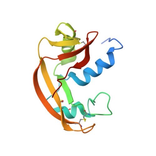
The widespread and functionally varied members of the ribonuclease A (RNase A) superfamily provide an excellent opportunity to study evolutionary forces at work on a conserved protein scaffold. Representatives from the zebrafish are of particular interest as the evolutionary distance from non-ichthyic homologues is large. We conducted an exhaustive survey of available zebrafish DNA sequences and found significant polymorphism among its four known homologues. In an extension of previous nomenclature, the variants have been named RNases ZF-1a-c,-2a-d,-3a-e and-4. We present the first X-ray crystal structures of zebrafish ribonucleases, RNases ZF-1a and-3e at 1.35-and 1.85 A resolution, respectively. Structure-based clustering with ten other ribonuclease structures indicates greatest similarity to mammalian angiogenins and amphibian ribonucleases, and supports the view that all present-day ribonucleases evolved from a progenitor with three disulphide bonds. In their details, the two structures are intriguing melting-pots of features present in ribonucleases from other vertebrate classes. Whereas in RNase ZF-1a the active site is obstructed by the C-terminal segment (as observed in angiogenin), in RNase ZF-3e the same region is open (as observed in more catalytically efficient homologues). The progenitor of present-day ribonucleases is more likely to have had an obstructive C terminus, and the relatively high similarity (late divergence) of RNases ZF-1 and-3 infers that the active site unblocking event has happened independently in different vertebrate lineages.
- Department of Biology and Biochemistry, University of Bath, Claverton Down, Bath BA2 7AY, UK.
Organizational Affiliation: