A New Series of N-[2,4-Dioxo-6-D-Ribitylamino-1,2, 3,4-Tetrahydropyrimidin-5-Yl]Oxalamic Acid Derivatives as Inhibitors of Lumazine Syntase and Riboflavin Synthase: Design, Synthesis, Biochemical Evaluation, Crystallography and Mechanistic Implications.
Zhang, Y., Illarionov, B., Morgunova, E., Bacher, A., Fischer, M., Ladenstein, R., Cushman, M.(2008) J Org Chem 73: 2715
- PubMed: 18331058 Search on PubMed
- DOI: https://doi.org/10.1021/jo702631a
- Primary Citation Related Structures:
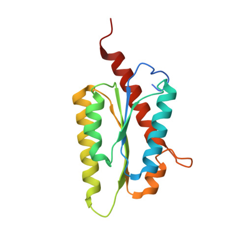
2VI5 - PubMed Abstract:
The penultimate step in the biosynthesis of riboflavin is catalyzed by lumazine synthase. Three metabolically stable analogues of the hypothetical intermediate proposed to arise after phosphate elimination in the lumazine synthase-catalyzed reaction were synthesized and evaluated as lumazine synthase inhibitors. All three intermediate analogues were inhibitors of Mycobacterium tuberculosis lumazine synthase, Bacillus subtilis lumazine synthase, and Schizosaccharomyces pombe lumazine synthase, while one of them proved to be an extremely potent inhibitor of Escherichia coli riboflavin synthase with a Ki of 1.3 nM. The crystal structure of M. tuberculosis lumazine synthase in complex with one of the inhibitors provides a model of the conformation of the intermediate occurring immediately after phosphate elimination, supporting a mechanism in which phosphate elimination occurs before a conformational change of the Schiff base intermediate toward a cyclic structure.
- The Purdue Cancer Center, Purdue University, West Lafayette, IN 47907, USA.
Organizational Affiliation: