Structure Determination and Biochemical Studies on Bacillus Stearothermophilus E53Q Serine Hydroxymethyltransferase and its Complexes Provide Insights on Function and Enzyme Memory
Rajaram, V., Bhavani, B.S., Kaul, P., Prakash, V., Appaji Rao, N., Savithri, H.S., Murthy, M.R.N.(2007) FEBS J 274: 4148
- PubMed: 17651438 Search on PubMed
- DOI: https://doi.org/10.1111/j.1742-4658.2007.05943.x
- Primary Citation Related Structures:
2VGS, 2VGT, 2VGU, 2VGV, 2VGW - PubMed Abstract:
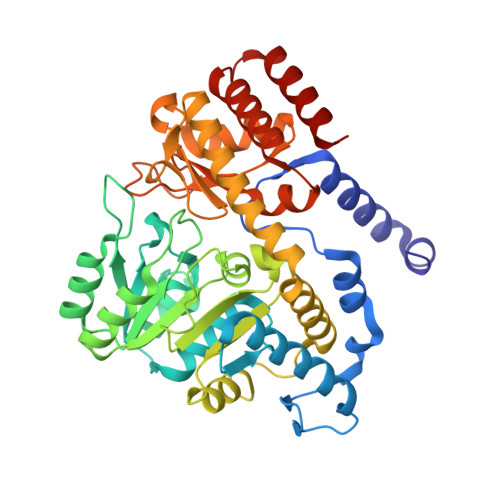
Serine hydroxymethyltransferase (SHMT) belongs to the alpha-family of pyridoxal 5'-phosphate-dependent enzymes and catalyzes the reversible conversion of L-Ser and tetrahydrofolate to Gly and 5,10-methylene tetrahydrofolate. 5,10-Methylene tetrahydrofolate serves as a source of one-carbon fragment in many biological processes. SHMT also catalyzes the tetrahydrofolate-independent conversion of L-allo-Thr to Gly and acetaldehyde. The crystal structure of Bacillus stearothermophilus SHMT (bsSHMT) suggested that E53 interacts with the substrate, L-Ser and tetrahydrofolate. To elucidate the role of E53, it was mutated to Q and structural and biochemical studies were carried out with the mutant enzyme. The internal aldimine structure of E53QbsSHMT was similar to that of the wild-type enzyme, except for significant changes at Q53, Y60 and Y61. The carboxyl of Gly and side chain of L-Ser were in two conformations in the respective external aldimine structures. The mutant enzyme was completely inactive for tetrahydrofolate-dependent cleavage of L-Ser, whereas there was a 1.5-fold increase in the rate of tetrahydrofolate-independent reaction with L-allo-Thr. The results obtained from these studies suggest that E53 plays an essential role in tetrahydrofolate/5-formyl tetrahydrofolate binding and in the proper positioning of Cbeta of L-Ser for direct attack by N5 of tetrahydrofolate. Most interestingly, the structure of the complex obtained by cocrystallization of E53QbsSHMT with Gly and 5-formyl tetrahydrofolate revealed the gem-diamine form of pyridoxal 5'-phosphate bound to Gly and active site Lys. However, density for 5-formyl tetrahydrofolate was not observed. Gly carboxylate was in a single conformation, whereas pyridoxal 5'-phosphate had two distinct conformations. The differences between the structures of this complex and Gly external aldimine suggest that the changes induced by initial binding of 5-formyl tetrahydrofolate are retained even though 5-formyl tetrahydrofolate is absent in the final structure. Spectral studies carried out with this mutant enzyme also suggest that 5-formyl tetrahydrofolate binds to the E53QbsSHMT-Gly complex forming a quinonoid intermediate and falls off within 4 h of dialysis, leaving behind the mutant enzyme in the gem-diamine form. This is the first report to provide direct evidence for enzyme memory based on the crystal structure of enzyme complexes.
- Molecular Biophysics Unit, Indian Institute of Science, Bangalore, India.
Organizational Affiliation: