2VGO | pdb_00002vgo

Crystal structure of Aurora B kinase in complex with Reversine inhibitor


Experimental Data Snapshot

  • Method: X-RAY DIFFRACTION
  • Resolution: 1.70 Å
  • R-Value Free: 
    0.229 (Depositor) 
  • R-Value Work: 
    0.197 (Depositor) 
  • R-Value Observed: 
    0.199 (Depositor) 

Starting Model: experimental
View more details

wwPDB Validation 3D Report Full Report

Validation slider image for 2VGO

Ligand Structure Quality Assessment 


This is version 2.2 of the entry. See complete history

Literature

Reversine, a Novel Aurora Kinases Inhibitor, Inhibits Colony Formation of Human Acute Myeloid Leukemia Cells.

D'Alise, A.M.Amabile, G.Iovino, M.Di Giorgio, F.P.Bartiromo, M.Sessa, F.Villa, F.Musacchio, A.Cortese, R.

(2008) Mol Cancer Ther 7: 1140

  • DOI: https://doi.org/10.1158/1535-7163.MCT-07-2051
  • Primary Citation Related Structures: 
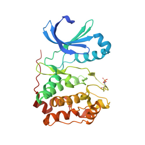
    2VGO

  • PubMed Abstract: 

    The demonstration that the small synthetic molecule reversine [2-(4-morpholinoanilino)-N6-cyclohexyladenine] promotes the dedifferentiation of committed cells into multipotent progenitor-type cells has raised hopes on the exploitation of this small chemical tool for the generation of stem cells. Here, we show that reversine causes a failure in cytokinesis and induces polyploidization. These effects of reversine are due to the inhibition of Aurora A and B, two related kinases that are implicated in several aspects of mitosis and that are frequently amplified and overexpressed in human tumors. Reversine inhibits the phosphorylation of histone H3, a direct downstream target of Aurora kinases. Similarly to the Aurora kinase inhibitor VX-680, which has recently entered phase II clinical trials for cancer treatment, reversine inhibited colony formation of leukemic cells from patients with acute myeloid leukemia but was significantly less toxic than VX-680 on cells from healthy donors. The crystal structure of the reversine-Aurora B kinase complex shows that reversine is a novel class of ATP-competitive Aurora kinase inhibitors. Thus, although our studies raise serious doubts on the application of reversine in regenerative medicine, they support the paradigm that reversine might be a useful agent in cancer chemotherapy.


  • Organizational Affiliation
    • CEINGE, Biotecnologie Avanzate, Via Comunale Margherita 482, 80131 Naples, Italy.

Macromolecule Content 

  • Total Structure Weight: 78.01 kDa 
  • Atom Count: 5,956 
  • Modeled Residue Count: 631 
  • Deposited Residue Count: 656 
  • Unique protein chains: 2

Macromolecules

Find similar proteins by:|  3D Structure
Entity ID: 1
MoleculeChains  Sequence LengthOrganismDetailsImage
SERINE/THREONINE-PROTEIN KINASE 12-A
A, B
284Xenopus laevisMutation(s): 0 
EC: 2.7.11.1
UniProt
Find proteins for Q6DE08 (Xenopus laevis)
Explore Q6DE08 
Go to UniProtKB:  Q6DE08
Entity Groups
Sequence Clusters30% Identity50% Identity70% Identity90% Identity95% Identity100% Identity
UniProt GroupQ6DE08
Sequence Annotations
Expand
Reference Sequence
Find similar proteins by:|  3D Structure
Entity ID: 2
MoleculeChains  Sequence LengthOrganismDetailsImage
INNER CENTROMERE PROTEIN A
C, D
44Xenopus laevisMutation(s): 0 
UniProt
Find proteins for O13024 (Xenopus laevis)
Explore O13024 
Go to UniProtKB:  O13024
Entity Groups
Sequence Clusters30% Identity50% Identity70% Identity90% Identity95% Identity100% Identity
UniProt GroupO13024
Sequence Annotations
Expand
Reference Sequence

Small Molecules

Ligands 1 Unique
IDChains Name / Formula / InChI Key2D Diagram3D Interactions
AD5

Query on AD5


Download:Ideal Coordinates CCD File
E [auth A],
F [auth B]
N~6~-cyclohexyl-N~2~-(4-morpholin-4-ylphenyl)-9H-purine-2,6-diamine
C21 H27 N7 O
ZFLJHSQHILSNCM-UHFFFAOYSA-N
Modified Residues  1 Unique
IDChains TypeFormula2D DiagramParent
TPO
Query on TPO
A, B
L-PEPTIDE LINKINGC4 H10 N O6 PTHR

Experimental Data & Validation

Experimental Data

  • Method: X-RAY DIFFRACTION
  • Resolution: 1.70 Å
  • R-Value Free:  0.229 (Depositor) 
  • R-Value Work:  0.197 (Depositor) 
  • R-Value Observed: 0.199 (Depositor) 
Space Group: P 1 21 1
Unit Cell:
Length ( Å )Angle ( ˚ )
a = 45.736α = 90
b = 67.6β = 96.59
c = 116.631γ = 90
Software Package:
Software NamePurpose
REFMACrefinement
HKL-2000data reduction
SCALEPACKdata scaling
MOLREPphasing

Structure Validation

View Full Validation Report


Ligand Structure Quality Assessment 


Entry History 

Deposition Data

Revision History  (Full details and data files)

  • Version 1.0: 2008-10-28
    Type: Initial release
  • Version 1.1: 2011-05-08
    Changes: Version format compliance
  • Version 1.2: 2011-07-13
    Changes: Version format compliance
  • Version 2.0: 2022-11-02
    Changes: Atomic model, Database references, Derived calculations, Other, Structure summary
  • Version 2.1: 2024-01-31
    Changes: Data collection, Refinement description
  • Version 2.2: 2024-10-23
    Changes: Structure summary