2VF9

Crystal structure of bacteriophage PRR1


Experimental Data Snapshot

  • Method: X-RAY DIFFRACTION
  • Resolution: 3.50 Å
  • R-Value Free: 0.313 
  • R-Value Work: 0.311 
  • R-Value Observed: 0.311 

wwPDB Validation   3D Report Full Report


This is version 1.2 of the entry. See complete history


Literature

The Capsid of the Small RNA Phage Prr1 is Stabilized by Metal Ions

Persson, M.Tars, K.Liljas, L.

(2008) J Mol Biol 383: 914

  • DOI: https://doi.org/10.1016/j.jmb.2008.08.060
  • Primary Citation of Related Structures:  
    2VF9

  • PubMed Abstract: 

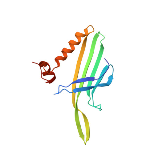
    Many nonenveloped virus particles are stabilized by calcium ions bound in the interfaces between the protein subunits. These ions may have a role in the disassembly process. The small RNA phages of the Leviviridae family have T=3 quasi-symmetry and are unique among simple viruses in that they have a coat protein with a translational repressor activity and a fold that has not been observed in other viruses. The crystal structure of phage PRR1 has been determined to 3.5 A resolution. The structure shows a tentative binding site for a calcium ion close to the quasi-3-fold axis. The RNA-binding surface used for repressor activity is mostly conserved. The structure does not show any significant differences between quasi-equivalent subunits, which suggests that the assembly is not controlled by conformational switches as in many other simple viruses.


  • Organizational Affiliation

    Department of Cell and Molecular Biology, Uppsala University, BMC, Husargatan 3, Box 596, S-751 24 Uppsala, Sweden.


Macromolecules
Find similar proteins by:  (by identity cutoff)  |  3D Structure
Entity ID: 1
MoleculeChains Sequence LengthOrganismDetailsImage
COAT PROTEIN
A, B, C
131Pseudomonas phage PRR1Mutation(s): 0 
UniProt
Find proteins for P03616 (Pseudomonas phage PRR1)
Explore P03616 
Go to UniProtKB:  P03616
Entity Groups  
Sequence Clusters30% Identity50% Identity70% Identity90% Identity95% Identity100% Identity
UniProt GroupP03616
Sequence Annotations
Expand
  • Reference Sequence
Small Molecules
Ligands 1 Unique
IDChains Name / Formula / InChI Key2D Diagram3D Interactions
CA
Query on CA

Download Ideal Coordinates CCD File 
D [auth C]CALCIUM ION
Ca
BHPQYMZQTOCNFJ-UHFFFAOYSA-N
Experimental Data & Validation

Experimental Data

  • Method: X-RAY DIFFRACTION
  • Resolution: 3.50 Å
  • R-Value Free: 0.313 
  • R-Value Work: 0.311 
  • R-Value Observed: 0.311 
  • Space Group: P 1
Unit Cell:
Length ( Å )Angle ( ˚ )
a = 281.4α = 93.27
b = 285.4β = 90
c = 472.5γ = 119.44
Software Package:
Software NamePurpose
CNSmodel building
PHASERmodel building
DENZOdata reduction
DENZOphasing
SCALEPACKphasing
GLRFphasing
CNSphasing
MOLEMAN2phasing
AVEphasing
Ophasing
PHASERphasing
MAMAphasing

Structure Validation

View Full Validation Report


Entry History 

Deposition Data

Revision History  (Full details and data files)

  • Version 1.0: 2008-09-23
    Type: Initial release
  • Version 1.1: 2012-02-01
    Changes: Other, Version format compliance
  • Version 1.2: 2023-12-13
    Changes: Data collection, Database references, Derived calculations, Other, Refinement description