2VD4

Structure of small-molecule inhibitor of Glmu from Haemophilus influenzae reveals an allosteric binding site


Experimental Data Snapshot

  • Method: X-RAY DIFFRACTION
  • Resolution: 1.90 Å
  • R-Value Free: 0.204 
  • R-Value Work: 0.182 
  • R-Value Observed: 0.183 

wwPDB Validation   3D Report Full Report


Ligand Structure Quality Assessment 


This is version 1.2 of the entry. See complete history


Literature

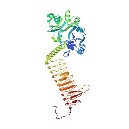
Structure of a Small-Molecule Inhibitor Complexed with Glmu from Haemophilus Influenzae Reveals an Allosteric Binding Site.

Mochalkin, I.Lightle, S.Narasimhan, L.Bornemeier, D.Melnick, M.Vanderroest, S.Mcdowell, L.

(2008) Protein Sci 17: 577

  • DOI: https://doi.org/10.1110/ps.073271408
  • Primary Citation of Related Structures:  
    2VD4

  • PubMed Abstract: 

    N-Acetylglucosamine-1-phosphate uridyltransferase (GlmU) is an essential enzyme in aminosugars metabolism and an attractive target for antibiotic drug discovery. GlmU catalyzes the formation of uridine-diphospho-N-acetylglucosamine (UDP-GlcNAc), an important precursor in the peptidoglycan and lipopolisaccharide biosynthesis in both Gram-negative and Gram-positive bacteria. Here we disclose a 1.9 A resolution crystal structure of a synthetic small-molecule inhibitor of GlmU from Haemophilus influenzae (hiGlmU). The compound was identified through a high-throughput screening (HTS) configured to detect inhibitors that target the uridyltransferase active site of hiGlmU. The original HTS hit exhibited a modest micromolar potency (IC(50) approximately 18 microM in a racemic mixture) against hiGlmU and no activity against Staphylococcus aureus GlmU (saGlmU). The determined crystal structure indicated that the inhibitor occupies an allosteric site adjacent to the GlcNAc-1-P substrate-binding region. Analysis of the mechanistic model of the uridyltransferase reaction suggests that the binding of this allosteric inhibitor prevents structural rearrangements that are required for the enzymatic reaction, thus providing a basis for structure-guided design of a new class of mechanism-based inhibitors of GlmU.


  • Organizational Affiliation

    Pfizer Inc., Michigan Laboratories, Ann Arbor, Michigan 48105, USA. Igor.Mochalkin@pfizer.com


Macromolecules
Find similar proteins by:  (by identity cutoff)  |  3D Structure
Entity ID: 1
MoleculeChains Sequence LengthOrganismDetailsImage
BIFUNCTIONAL PROTEIN GLMU456Haemophilus influenzaeMutation(s): 0 
EC: 2
UniProt
Find proteins for P43889 (Haemophilus influenzae (strain ATCC 51907 / DSM 11121 / KW20 / Rd))
Explore P43889 
Go to UniProtKB:  P43889
Entity Groups  
Sequence Clusters30% Identity50% Identity70% Identity90% Identity95% Identity100% Identity
UniProt GroupP43889
Sequence Annotations
Expand
  • Reference Sequence
Small Molecules
Ligands 5 Unique
IDChains Name / Formula / InChI Key2D Diagram3D Interactions
P21
Query on P21

Download Ideal Coordinates CCD File 
B [auth A]4-chloro-N-(3-methoxypropyl)-N-[(3S)-1-(2-phenylethyl)piperidin-3-yl]benzamide
C24 H31 Cl N2 O2
OOUGHDJEJNMXSV-QHCPKHFHSA-N
PG4
Query on PG4

Download Ideal Coordinates CCD File 
H [auth A],
I [auth A]
TETRAETHYLENE GLYCOL
C8 H18 O5
UWHCKJMYHZGTIT-UHFFFAOYSA-N
PGE
Query on PGE

Download Ideal Coordinates CCD File 
J [auth A]TRIETHYLENE GLYCOL
C6 H14 O4
ZIBGPFATKBEMQZ-UHFFFAOYSA-N
SO4
Query on SO4

Download Ideal Coordinates CCD File 
C [auth A]
D [auth A]
E [auth A]
F [auth A]
G [auth A]
C [auth A],
D [auth A],
E [auth A],
F [auth A],
G [auth A],
M [auth A]
SULFATE ION
O4 S
QAOWNCQODCNURD-UHFFFAOYSA-L
MG
Query on MG

Download Ideal Coordinates CCD File 
K [auth A],
L [auth A]
MAGNESIUM ION
Mg
JLVVSXFLKOJNIY-UHFFFAOYSA-N
Binding Affinity Annotations 
IDSourceBinding Affinity
P21 Binding MOAD:  2VD4 IC50: 1.80e+4 (nM) from 1 assay(s)
PDBBind:  2VD4 IC50: 1.80e+4 (nM) from 1 assay(s)
Experimental Data & Validation

Experimental Data

  • Method: X-RAY DIFFRACTION
  • Resolution: 1.90 Å
  • R-Value Free: 0.204 
  • R-Value Work: 0.182 
  • R-Value Observed: 0.183 
  • Space Group: H 3 2
Unit Cell:
Length ( Å )Angle ( ˚ )
a = 108.186α = 90
b = 108.186β = 90
c = 327.43γ = 120
Software Package:
Software NamePurpose
HKL-2000data reduction
HKL-2000data scaling
CNXphasing
REFMACphasing
REFMACrefinement

Structure Validation

View Full Validation Report


Ligand Structure Quality Assessment 


Entry History 

Deposition Data

Revision History  (Full details and data files)

  • Version 1.0: 2008-01-15
    Type: Initial release
  • Version 1.1: 2011-07-13
    Changes: Advisory, Version format compliance
  • Version 1.2: 2023-12-13
    Changes: Data collection, Database references, Derived calculations, Other, Refinement description