


The Crystal Structure of Hisg from B. Subtilis
Lohkamp, B., Riboldi-Tunniclife, A., Lapthorn, A.J.To be published.
Experimental Data Snapshot
wwPDB Validation 3D Report Full Report

Entity ID: 1 | |||||
|---|---|---|---|---|---|
| Molecule | Chains | Sequence Length | Organism | Details | Image |
| ATP PHOSPHORIBOSYLTRANSFERASE | 214 | Bacillus subtilis | Mutation(s): 0 EC: 2.4.2.17 | 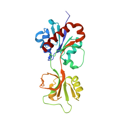 | |
UniProt | |||||
Entity Groups | |||||
| Sequence Clusters | 30% Identity50% Identity70% Identity90% Identity95% Identity100% Identity | ||||
| UniProt Group | O34520 | ||||
Sequence AnnotationsExpand | |||||
Reference Sequence | |||||
| Length ( Å ) | Angle ( ˚ ) |
|---|---|
| a = 85.937 | α = 90 |
| b = 85.937 | β = 90 |
| c = 75.948 | γ = 90 |
| Software Name | Purpose |
|---|---|
| REFMAC | refinement |


