Intervening with Urinary Tract Infections Using Anti-Adhesives Based on the Crystal Structure of the Fimh-Oligomannose-3 Complex.
Wellens, A., Garofalo, C., Nguyen, H., Van Gerven, N., Slattegard, R., Hernalsteens, J.P., Wyns, L., Oscarson, S., De Greve, H., Hultgren, S.J., Bouckaert, J.(2008) PLoS One 3: E2040
- PubMed: 18446213 Search on PubMedSearch on PubMed Central
- DOI: https://doi.org/10.1371/journal.pone.0002040
- Primary Citation Related Structures:
2VCO - PubMed Abstract:
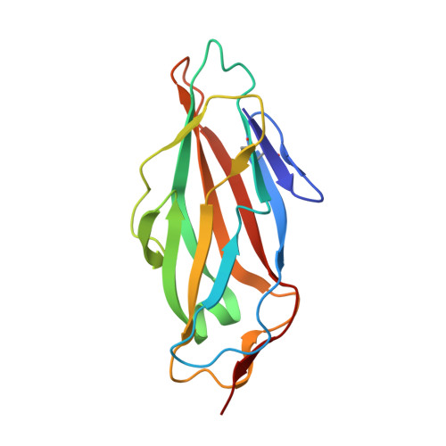
Escherichia coli strains adhere to the normally sterile human uroepithelium using type 1 pili, that are long, hairy surface organelles exposing a mannose-binding FimH adhesin at the tip. A small percentage of adhered bacteria can successfully invade bladder cells, presumably via pathways mediated by the high-mannosylated uroplakin-Ia and alpha3beta1 integrins found throughout the uroepithelium. Invaded bacteria replicate and mature into dense, biofilm-like inclusions in preparation of fluxing and of infection of neighbouring cells, being the major cause of the troublesome recurrent urinary tract infections. We demonstrate that alpha-D-mannose based inhibitors of FimH not only block bacterial adhesion on uroepithelial cells but also antagonize invasion and biofilm formation. Heptyl alpha-D-mannose prevents binding of type 1-piliated E. coli to the human bladder cell line 5637 and reduces both adhesion and invasion of the UTI89 cystitis isolate instilled in mouse bladder via catheterization. Heptyl alpha-D-mannose also specifically inhibited biofilm formation at micromolar concentrations. The structural basis of the great inhibitory potential of alkyl and aryl alpha-D-mannosides was elucidated in the crystal structure of the FimH receptor-binding domain in complex with oligomannose-3. FimH interacts with Man alpha1,3Man beta1,4GlcNAc beta1,4GlcNAc in an extended binding site. The interactions along the alpha1,3 glycosidic bond and the first beta1,4 linkage to the chitobiose unit are conserved with those of FimH with butyl alpha-D-mannose. The strong stacking of the central mannose with the aromatic ring of Tyr48 is congruent with the high affinity found for synthetic inhibitors in which this mannose is substituted for by an aromatic group. The potential of ligand-based design of antagonists of urinary tract infections is ruled by the structural mimicry of natural epitopes and extends into blocking of bacterial invasion, intracellular growth and capacity to fluxing and of recurrence of the infection.
- Department of Molecular and Cellular Interactions, VIB [corrected] Brussels, Belgium.
Organizational Affiliation: