Molecular model of a type III secretion system needle: Implications for host-cell sensing.
Deane, J.E., Roversi, P., Cordes, F.S., Johnson, S., Kenjale, R., Daniell, S., Booy, F., Picking, W.D., Picking, W.L., Blocker, A.J., Lea, S.M.(2006) Proc Natl Acad Sci U S A 103: 12529-12533
- PubMed: 16888041 Search on PubMedSearch on PubMed Central
- DOI: https://doi.org/10.1073/pnas.0602689103
- Primary Citation Related Structures:
2CA5, 2V6L - PubMed Abstract:
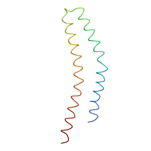
Type III secretion systems are essential virulence determinants for many Gram-negative bacterial pathogens. The type III secretion system consists of cytoplasmic, transmembrane, and extracellular domains. The extracellular domain is a hollow needle protruding above the bacterial surface and is held within a basal body that traverses both bacterial membranes. Effector proteins are translocated, via this external needle, directly into host cells, where they subvert normal cell functions to aid infection. Physical contact with host cells initiates secretion and leads to formation of a pore, thought to be contiguous with the needle channel, in the host-cell membrane. Here, we report the crystal structure of the Shigella flexneri needle subunit MxiH and a complete model for the needle assembly built into our three-dimensional EM reconstruction. The model, combined with mutagenesis data, reveals that signaling of host-cell contact is relayed through the needle via intersubunit contacts and suggests a mode of binding for a tip complex.
- Laboratory of Molecular Biophysics, Department of Biochemistry, University of Oxford, Oxford OX1 3QU, United Kingdom.
Organizational Affiliation: