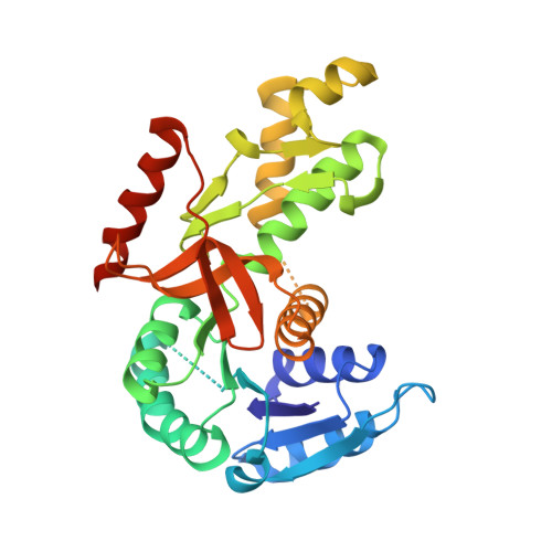
Activity, stability and structural studies of lactate dehydrogenases adapted to extreme thermal environments.
Coquelle, N., Fioravanti, E., Weik, M., Vellieux, F., Madern, D.(2007) J Mol Biology 374: 547-562
- PubMed: 17936781
- DOI: https://doi.org/10.1016/j.jmb.2007.09.049
- Primary Citation of Related Structures:
2V65, 2V6B, 2V6M, 2V7P - PubMed Abstract:
Lactate dehydrogenase (LDH) catalyzes the conversion of pyruvate to lactate with concomitant oxidation of NADH during the last step in anaerobic glycolysis. In the present study, we present a comparative biochemical and structural analysis of various LDHs adapted to function over a large temperature range. The enzymes were from Champsocephalus gunnari (an Antarctic fish), Deinococcus radiodurans (a mesophilic bacterium) and Thermus thermophilus (a hyperthermophilic bacterium). The thermodynamic activation parameters of these LDHs indicated that temperature adaptation from hot to cold conditions was due to a decrease in the activation enthalpy and an increase in activation entropy. The crystal structures of these LDHs have been solved. Pairwise comparisons at the structural level, between hyperthermophilic versus mesophilic LDHs and mesophilic versus psychrophilic LDHs, have revealed that temperature adaptation is due to a few amino acid substitutions that are localized in critical regions of the enzyme. These substitutions, each having accumulating effects, play a role in either the conformational stability or the local flexibility or in both. Going from hot- to cold-adapted LDHs, the various substitutions have decreased the number of ion pairs, reduced the size of ionic networks, created unfavorable interactions involving charged residues and induced strong local disorder. The analysis of the LDHs adapted to extreme temperatures shed light on how evolutionary processes shift the subtle balance between overall stability and flexibility of an enzyme.
- Laboratoire de Biophysique Moléculaire, Institut de Biologie Structurale J.-P. Ebel, CEA CNRS UJF, UMR 5075, 41 rue Jules Horowitz, 38027 Grenoble Cedex 01, France.
Organizational Affiliation:


