The Structure of the Coiled-Coil Domain of Ndel1 and the Basis of its Interaction with Lis1, the Causal Protein of Miller-Dieker Lissencephaly.
Derewenda, U., Tarricone, C., Choi, W.C., Cooper, D.R., Lukasik, S., Perrina, F., Tripathy, A., Kim, M.H., Cafiso, D.S., Musacchio, A., Derewenda, Z.S.(2007) Structure 15: 1467
- PubMed: 17997972 Search on PubMed
- DOI: https://doi.org/10.1016/j.str.2007.09.015
- Primary Citation Related Structures:
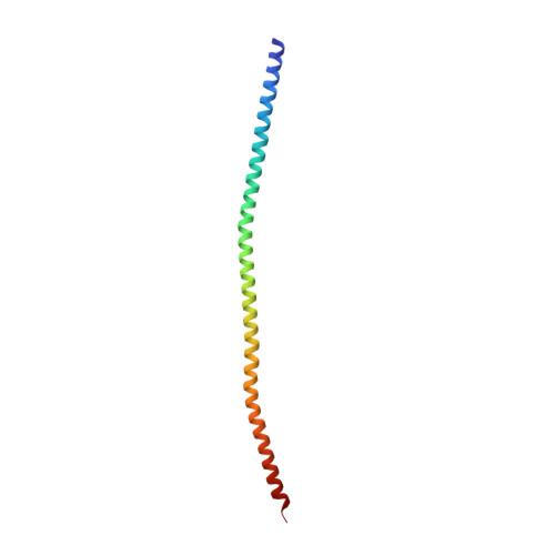
2V66, 2V71 - PubMed Abstract:
Ndel1 and Nde1 are homologous and evolutionarily conserved proteins, with critical roles in cell division, neuronal migration, and other physiological phenomena. These functions are dependent on their interactions with the retrograde microtubule motor dynein and with its regulator Lis1--a product of the causal gene for isolated lissencephaly sequence (ILS) and Miller-Dieker lissencephaly. The molecular basis of the interactions of Ndel1 and Nde1 with Lis1 is not known. Here, we present a crystallographic study of two fragments of the coiled-coil domain of Ndel1, one of which reveals contiguous high-quality electron density for residues 10-166, the longest such structure reported by X-ray diffraction at high resolution. Together with complementary solution studies, our structures reveal how the Ndel1 coiled coil forms a stable parallel homodimer and suggest mechanisms by which the Lis1-interacting domain can be regulated to maintain a conformation in which two supercoiled alpha helices cooperatively bind to a Lis1 homodimer.
- Department of Molecular Physiology and Biological Physics, University of Virginia, Charlottesville, VA 22908, USA.
Organizational Affiliation: