The Crystal Structure of Fdxa, a 7Fe Ferredoxin from Mycobacterium Smegmatis.
Ricagno, S., De Rosa, M., Aliverti, A., Zanetti, G., Bolognesi, M.(2007) Biochem Biophys Res Commun 360: 97
- PubMed: 17577575 Search on PubMed
- DOI: https://doi.org/10.1016/j.bbrc.2007.06.013
- Primary Citation Related Structures:
2V2K - PubMed Abstract:
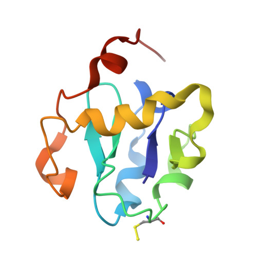
Mycobacterium smegmatis ferredoxin FdxA, which has an orthologue ferredoxin in Mycobacterium tuberculosis, FdxC, contains both one [3Fe-4S] and one [4Fe-4S] cluster. M. smegmatis FdxA has been shown to be a preferred ferredoxin substrate of FprA [F. Fischer, D. Raimondi, A. Aliverti, G. Zanetti, Mycobacterium tuberculosis FprA, a novel bacterial NADPH-ferredoxin reductase, Eur. J. Biochem. 269 (2002) 3005-3013], an adrenodoxin reductase-like flavoprotein of M. tuberculosis, suggesting that M. tuberculosis FdxC could be the physiological partner of the enzyme in providing reducing power to the cytochromes P450. We report here the crystal structure of FdxA at 1.6A resolution (R(factor) 16.5%, R(free) 20.2%). Besides providing an insight on protein architecture for this 106-residue ferredoxin, our crystallographic investigation highlights lability of the [4Fe-4S] center, which is shown to loose a Fe atom during crystal growth. Due to their high similarity (87% sequence identity), the structure here reported can be considered a valuable model for M. tuberculosis FdxC, thus representing a step forward in the study of the complex mycobacterial redox pathways.
- Department of Biomolecular Sciences and Biotechnology, University of Milano, Via Celoria 26, 20133 Milano, Italy.
Organizational Affiliation: