2V0S | pdb_00002v0s

crystal structure of a hairpin exchange variant (LR1) of the targeting LINE-1 retrotransposon endonuclease


Experimental Data Snapshot

  • Method: X-RAY DIFFRACTION
  • Resolution: 1.80 Å
  • R-Value Free: 
    0.226 (Depositor), 0.236 (DCC) 
  • R-Value Work: 
    0.188 (Depositor), 0.207 (DCC) 
  • R-Value Observed: 
    0.190 (Depositor) 

Starting Model: experimental
View more details

wwPDB Validation 3D Report Full Report

Validation slider image for 2V0S

This is version 1.3 of the entry. See complete history

Literature

Determinants for DNA Target Structure Selectivity of the Human Line-1 Retrotransposon Endonuclease.

Repanas, K.Zingler, N.Layer, L.E.Schumann, G.G.Perrakis, A.Weichenrieder, O.

(2007) Nucleic Acids Res 35: 4914

  • DOI: https://doi.org/10.1093/nar/gkm516
  • Primary Citation Related Structures: 
    2V0R, 2V0S

  • PubMed Abstract: 

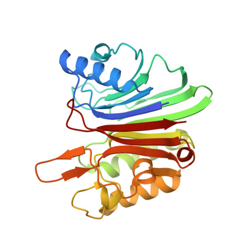
    The human LINE-1 endonuclease (L1-EN) is the targeting endonuclease encoded by the human LINE-1 (L1) retrotransposon. L1-EN guides the genomic integration of new L1 and Alu elements that presently account for approximately 28% of the human genome. L1-EN bears considerable technological interest, because its target selectivity may ultimately be engineered to allow the site-specific integration of DNA into defined genomic locations. Based on the crystal structure, we generated L1-EN mutants to analyze and manipulate DNA target site recognition. Crystal structures and their dynamic and functional analysis show entire loop grafts to be feasible, resulting in altered specificity, while individual point mutations do not change the nicking pattern of L1-EN. Structural parameters of the DNA target seem more important for recognition than the nucleotide sequence, and nicking profiles on DNA oligonucleotides in vitro are less well defined than the respective integration site consensus in vivo. This suggests that additional factors other than the DNA nicking specificity of L1-EN contribute to the targeted integration of non-LTR retrotransposons.


  • Organizational Affiliation
    • Division of Molecular Carcinogenesis, The Netherlands Cancer Institute, 1066 CX Amsterdam, The Netherlands.

Macromolecule Content 

  • Total Structure Weight: 27.11 kDa 
  • Atom Count: 2,022 
  • Modeled Residue Count: 229 
  • Deposited Residue Count: 236 
  • Unique protein chains: 1

Macromolecules

Find similar proteins by:|  3D Structure
Entity ID: 1
MoleculeChains  Sequence LengthOrganismDetailsImage
LR1236Homo sapiensMutation(s): 0 
EC: 2.7.7.49 (UniProt), 3.1.21 (UniProt)
UniProt
Find proteins for O00370 (Homo sapiens)
Explore O00370 
Go to UniProtKB:  O00370
Entity Groups
Sequence Clusters30% Identity50% Identity70% Identity90% Identity95% Identity100% Identity
UniProt GroupO00370
Sequence Annotations
Expand
Reference Sequence

Experimental Data & Validation

Experimental Data

  • Method: X-RAY DIFFRACTION
  • Resolution: 1.80 Å
  • R-Value Free:  0.226 (Depositor), 0.236 (DCC) 
  • R-Value Work:  0.188 (Depositor), 0.207 (DCC) 
  • R-Value Observed: 0.190 (Depositor) 
Space Group: C 2 2 21
Unit Cell:
Length ( Å )Angle ( ˚ )
a = 58.698α = 90
b = 67.648β = 90
c = 128.278γ = 90
Software Package:
Software NamePurpose
REFMACrefinement
MOSFLMdata reduction
SCALAdata scaling
MOLREPphasing

Structure Validation

View Full Validation Report


Entry History 

Deposition Data

Revision History  (Full details and data files)

  • Version 1.0: 2007-07-17
    Type: Initial release
  • Version 1.1: 2011-05-08
    Changes: Version format compliance
  • Version 1.2: 2011-07-13
    Changes: Version format compliance
  • Version 1.3: 2023-12-13
    Changes: Data collection, Database references, Derived calculations, Other, Refinement description