2V04

CRYSTAL STRUCTURE OF CHOLINE BINDING PROTEIN F FROM STREPTOCOCCUS PNEUMONIAE


Experimental Data Snapshot

  • Method: X-RAY DIFFRACTION
  • Resolution: 2.10 Å
  • R-Value Free: 0.250 
  • R-Value Work: 0.209 
  • R-Value Observed: 0.211 

wwPDB Validation   3D Report Full Report


This is version 1.2 of the entry. See complete history


Literature

Crystal Structure of Cbpf, a Bifunctional Choline-Binding Protein and Autolysis Regulator from Streptococcus Pneumoniae.

Molina, R.Gonzalez, A.Stelter, M.Perez-Dorado, I.Kahn, R.Morales, M.Campuzano, S.Campillo, N.E.Mobashery, S.Garcia, J.L.Garcia, P.Hermoso, J.A.

(2009) EMBO Rep 10: 246

  • DOI: https://doi.org/10.1038/embor.2008.245
  • Primary Citation of Related Structures:  
    2V04, 2VYU

  • PubMed Abstract: 

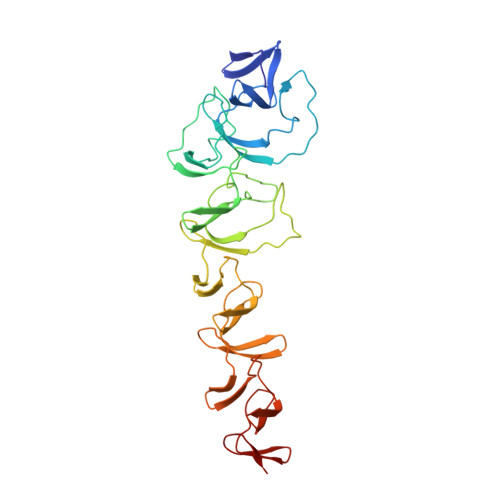
    Phosphorylcholine, a crucial component of the pneumococcal cell wall, is essential in bacterial physiology and in human pathogenesis because it binds to serum components of the immune system and acts as a docking station for the family of surface choline-binding proteins. The three-dimensional structure of choline-binding protein F (CbpF), one of the most abundant proteins in the pneumococcal cell wall, has been solved in complex with choline. CbpF shows a new modular structure composed both of consensus and non-consensus choline-binding repeats, distributed along its length, which markedly alter its shape, charge distribution and binding ability, and organizing the protein into two well-defined modules. The carboxy-terminal module is involved in cell wall binding and the amino-terminal module is crucial for inhibition of the autolytic LytC muramidase, providing a regulatory function for pneumococcal autolysis.


  • Organizational Affiliation

    Grupo de Cristalografia Macromolecular y Biologia Estructural, Instituto Química-Física Rocasolano, CSIC, Serrano 119, 28006 Madrid, Spain.


Macromolecules
Find similar proteins by:  (by identity cutoff)  |  3D Structure
Entity ID: 1
MoleculeChains Sequence LengthOrganismDetailsImage
CHOLINE BINDING PROTEIN F311Streptococcus pneumoniae R6Mutation(s): 0 
UniProt
Find proteins for Q8DR52 (Streptococcus pneumoniae (strain ATCC BAA-255 / R6))
Explore Q8DR52 
Go to UniProtKB:  Q8DR52
Entity Groups  
Sequence Clusters30% Identity50% Identity70% Identity90% Identity95% Identity100% Identity
UniProt GroupQ8DR52
Sequence Annotations
Expand
  • Reference Sequence
Experimental Data & Validation

Experimental Data

  • Method: X-RAY DIFFRACTION
  • Resolution: 2.10 Å
  • R-Value Free: 0.250 
  • R-Value Work: 0.209 
  • R-Value Observed: 0.211 
  • Space Group: P 21 21 2
Unit Cell:
Length ( Å )Angle ( ˚ )
a = 52.397α = 90
b = 115.792β = 90
c = 72.985γ = 90
Software Package:
Software NamePurpose
REFMACrefinement
MOSFLMdata reduction
SCALAdata scaling
SHELXphasing
SHARPphasing
MOLREPphasing

Structure Validation

View Full Validation Report


Entry History 

Deposition Data

Revision History  (Full details and data files)

  • Version 1.0: 2008-06-10
    Type: Initial release
  • Version 1.1: 2011-07-13
    Changes: Advisory, Refinement description, Version format compliance
  • Version 1.2: 2018-04-25
    Changes: Data collection, Database references