The Identity of the Active Site of Oxalate Decarboxylase and the Importance of the Stability of Active-Site Lid Conformations.
Just, V.J., Burrell, M.R., Bowater, L., Mcrobbie, I., Stevenson, C.E.M., Lawson, D.M., Bornemann, S.(2007) Biochem J 407: 397
- PubMed: 17680775 Search on PubMedSearch on PubMed Central
- DOI: https://doi.org/10.1042/BJ20070708
- Primary Citation Related Structures:
2UY8, 2UY9, 2UYA, 2UYB - PubMed Abstract:
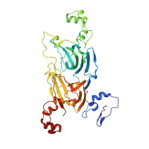
Oxalate decarboxylase (EC 4.1.1.2) catalyses the conversion of oxalate into carbon dioxide and formate. It requires manganese and, uniquely, dioxygen for catalysis. It forms a homohexamer and each subunit contains two similar, but distinct, manganese sites termed sites 1 and 2. There is kinetic evidence that only site 1 is catalytically active and that site 2 is purely structural. However, the kinetics of enzymes with mutations in site 2 are often ambiguous and all mutant kinetics have been interpreted without structural information. Nine new site-directed mutants have been generated and four mutant crystal structures have now been solved. Most mutants targeted (i) the flexibility (T165P), (ii) favoured conformation (S161A, S164A, D297A or H299A) or (iii) presence (Delta162-163 or Delta162-164) of a lid associated with site 1. The kinetics of these mutants were consistent with only site 1 being catalytically active. This was particularly striking with D297A and H299A because they disrupted hydrogen bonds between the lid and a neighbouring subunit only when in the open conformation and were distant from site 2. These observations also provided the first evidence that the flexibility and stability of lid conformations are important in catalysis. The deletion of the lid to mimic the plant oxalate oxidase led to a loss of decarboxylase activity, but only a slight elevation in the oxalate oxidase side reaction, implying other changes are required to afford a reaction specificity switch. The four mutant crystal structures (R92A, E162A, Delta162-163 and S161A) strongly support the hypothesis that site 2 is purely structural.
- Department of Biological Chemistry, John Innes Centre, Norwich Research Park, Norwich NR4 7UH, UK.
Organizational Affiliation: