2UVP

Crystal structure of HobA (HP1230)from Helicobacter pylori


Experimental Data Snapshot

  • Method: X-RAY DIFFRACTION
  • Resolution: 1.70 Å
  • R-Value Free: 0.214 
  • R-Value Work: 0.180 
  • R-Value Observed: 0.182 

wwPDB Validation   3D Report Full Report


This is version 1.2 of the entry. See complete history


Literature

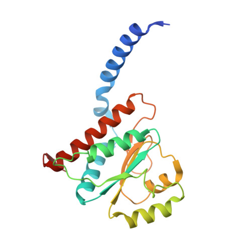
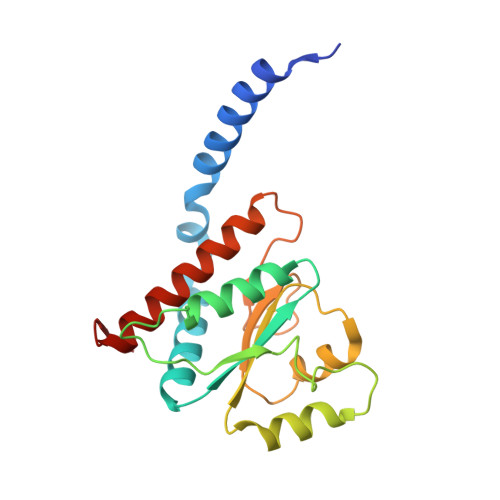
Structural similarity between the DnaA-binding proteins HobA (HP1230) from Helicobacter pylori and DiaA from Escherichia coli.

Natrajan, G.Hall, D.R.Thompson, A.C.Gutsche, I.Terradot, L.

(2007) Mol Microbiol 65: 995-1005

  • DOI: https://doi.org/10.1111/j.1365-2958.2007.05843.x
  • Primary Citation of Related Structures:  
    2UVP

  • PubMed Abstract: 

    In prokaryotes, DNA replication is initiated by the binding of DnaA to the oriC region of the chromosome to load the primosome machinery and start a new replication round. Several proteins control these events in Escherichia coli to ensure that replication is precisely timed during the cell cycle. Here, we report the crystal structure of HobA (HP1230) at 1.7 A, a recently discovered protein that specifically interacts with DnaA protein from Helicobacter pylori (HpDnaA). We found that the closest structural homologue of HobA is a sugar isomerase (SIS) domain containing protein, the phosphoheptose isomerase from Pseudomonas aeruginosa. Remarkably, SIS proteins share strong sequence homology with DiaA from E. coli; yet, HobA and DiaA share no sequence homology. Thus, by solving the structure of HobA, we unexpectedly discovered that HobA is a H. pylori structural homologue of DiaA. By comparing the structure of HobA to a homology model of DiaA, we identified conserved, surface-accessible residues that could be involved in protein-protein interaction. Finally, we show that HobA specifically interacts with the N-terminal part of HpDnaA. The structural homology between DiaA and HobA strongly supports their involvement in the replication process and these proteins could define a new structural family of replication regulators in bacteria.


  • Organizational Affiliation

    Macromolecular Crystallography Group, European Synchrotron Radiation Facility, B.P. 220, 6 rue Jules Horowitz, F-38043 Grenoble Cedex, France.


Macromolecules
Find similar proteins by:  (by identity cutoff)  |  3D Structure
Entity ID: 1
MoleculeChains Sequence LengthOrganismDetailsImage
HOBA186Helicobacter pyloriMutation(s): 0 
UniProt
Find proteins for O25828 (Helicobacter pylori (strain ATCC 700392 / 26695))
Explore O25828 
Go to UniProtKB:  O25828
Entity Groups  
Sequence Clusters30% Identity50% Identity70% Identity90% Identity95% Identity100% Identity
UniProt GroupO25828
Sequence Annotations
Expand
  • Reference Sequence
Find similar proteins by:  (by identity cutoff)  |  3D Structure
Entity ID: 2
MoleculeChains Sequence LengthOrganismDetailsImage
HOBA
B, C, D
186Helicobacter pyloriMutation(s): 0 
UniProt
Find proteins for O25828 (Helicobacter pylori (strain ATCC 700392 / 26695))
Explore O25828 
Go to UniProtKB:  O25828
Entity Groups  
Sequence Clusters30% Identity50% Identity70% Identity90% Identity95% Identity100% Identity
UniProt GroupO25828
Sequence Annotations
Expand
  • Reference Sequence
Small Molecules
Ligands 3 Unique
IDChains Name / Formula / InChI Key2D Diagram3D Interactions
GOL
Query on GOL

Download Ideal Coordinates CCD File 
H [auth B],
N [auth D]
GLYCEROL
C3 H8 O3
PEDCQBHIVMGVHV-UHFFFAOYSA-N
ACT
Query on ACT

Download Ideal Coordinates CCD File 
E [auth A],
F [auth A],
L [auth C]
ACETATE ION
C2 H3 O2
QTBSBXVTEAMEQO-UHFFFAOYSA-M
CA
Query on CA

Download Ideal Coordinates CCD File 
G [auth B],
I [auth B],
J [auth C],
K [auth C],
M [auth C]
CALCIUM ION
Ca
BHPQYMZQTOCNFJ-UHFFFAOYSA-N
Experimental Data & Validation

Experimental Data

  • Method: X-RAY DIFFRACTION
  • Resolution: 1.70 Å
  • R-Value Free: 0.214 
  • R-Value Work: 0.180 
  • R-Value Observed: 0.182 
  • Space Group: P 32
Unit Cell:
Length ( Å )Angle ( ˚ )
a = 112.232α = 90
b = 112.232β = 90
c = 61.929γ = 120
Software Package:
Software NamePurpose
REFMACrefinement
MOSFLMdata reduction
SCALAdata scaling
SHARPphasing

Structure Validation

View Full Validation Report


Entry History 

Deposition Data

Revision History  (Full details and data files)

  • Version 1.0: 2007-08-21
    Type: Initial release
  • Version 1.1: 2014-12-03
    Changes: Data collection, Derived calculations, Non-polymer description, Other, Structure summary, Version format compliance
  • Version 1.2: 2018-06-13
    Changes: Data collection, Database references, Source and taxonomy