2UV4 | pdb_00002uv4

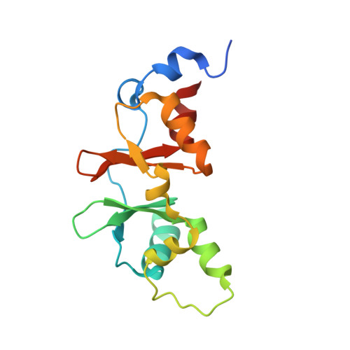
Crystal Structure of a CBS domain pair from the regulatory gamma1 subunit of human AMPK in complex with AMP


Experimental Data Snapshot

  • Method: X-RAY DIFFRACTION
  • Resolution: 1.33 Å
  • R-Value Free: 
    0.207 (Depositor) 
  • R-Value Work: 
    0.169 (Depositor) 
  • R-Value Observed: 
    0.171 (Depositor) 

wwPDB Validation 3D Report Full Report

Validation slider image for 2UV4

Ligand Structure Quality Assessment 


This is version 1.3 of the entry. See complete history. 

Literature

Structure of a Cbs-Domain Pair from the Regulatory Gamma1 Subunit of Human Ampk in Complex with AMP and Zmp.

Day, P., Sharff, A., Parra, L., Cleasby, A., Williams, M., Horer, S., Nar, H., Redemann, N., Tickle, I., Yon, J.

(2007) Acta Crystallogr D Biol Crystallogr 63: 587

  • DOI: https://doi.org/10.1107/S0907444907009110
  • Primary Citation Related Structures: 
    2UV4, 2UV5, 2UV6, 2UV7

  • PubMed Abstract: 

    AMP-activated kinase (AMPK) is central to sensing energy status in eukaryotic cells via binding of AMP and ATP to CBS (cystathionine beta-synthase) domains in the regulatory gamma subunit. The structure of a CBS-domain pair from human AMPK gamma1 in complex with the physiological activator AMP and the pharmacological activator ZMP (AICAR) is presented.


  • Organizational Affiliation: 
    • Astex Therapeutics Ltd, 436 Cambridge Science Park, Milton Road, Cambridge CB4 0QA, England. p.day@astex-therapeutics.com

Macromolecule Content 

  • Total Structure Weight: 17.63 kDa 
  • Atom Count: 1,416 
  • Modeled Residue Count: 144 
  • Deposited Residue Count: 152 
  • Unique protein chains: 1

Macromolecules

Find similar proteins by:|  3D Structure
Entity ID: 1
MoleculeChains  Sequence LengthOrganismDetailsImage
5'-AMP-ACTIVATED PROTEIN KINASE SUBUNIT GAMMA-1152Homo sapiensMutation(s): 0 
UniProt & NIH Common Fund Data Resources
Find proteins for P54619 (Homo sapiens)
Explore P54619 
Go to UniProtKB:  P54619
PHAROS:  P54619
GTEx:  ENSG00000181929 
Entity Groups
Sequence Clusters30% Identity50% Identity70% Identity90% Identity95% Identity100% Identity
UniProt GroupP54619
Sequence Annotations
Expand
Reference Sequence

Small Molecules

Ligands 1 Unique
IDChains Name / Formula / InChI Key2D Diagram3D Interactions
AMP

Query on AMP


Download:Ideal Coordinates CCD File
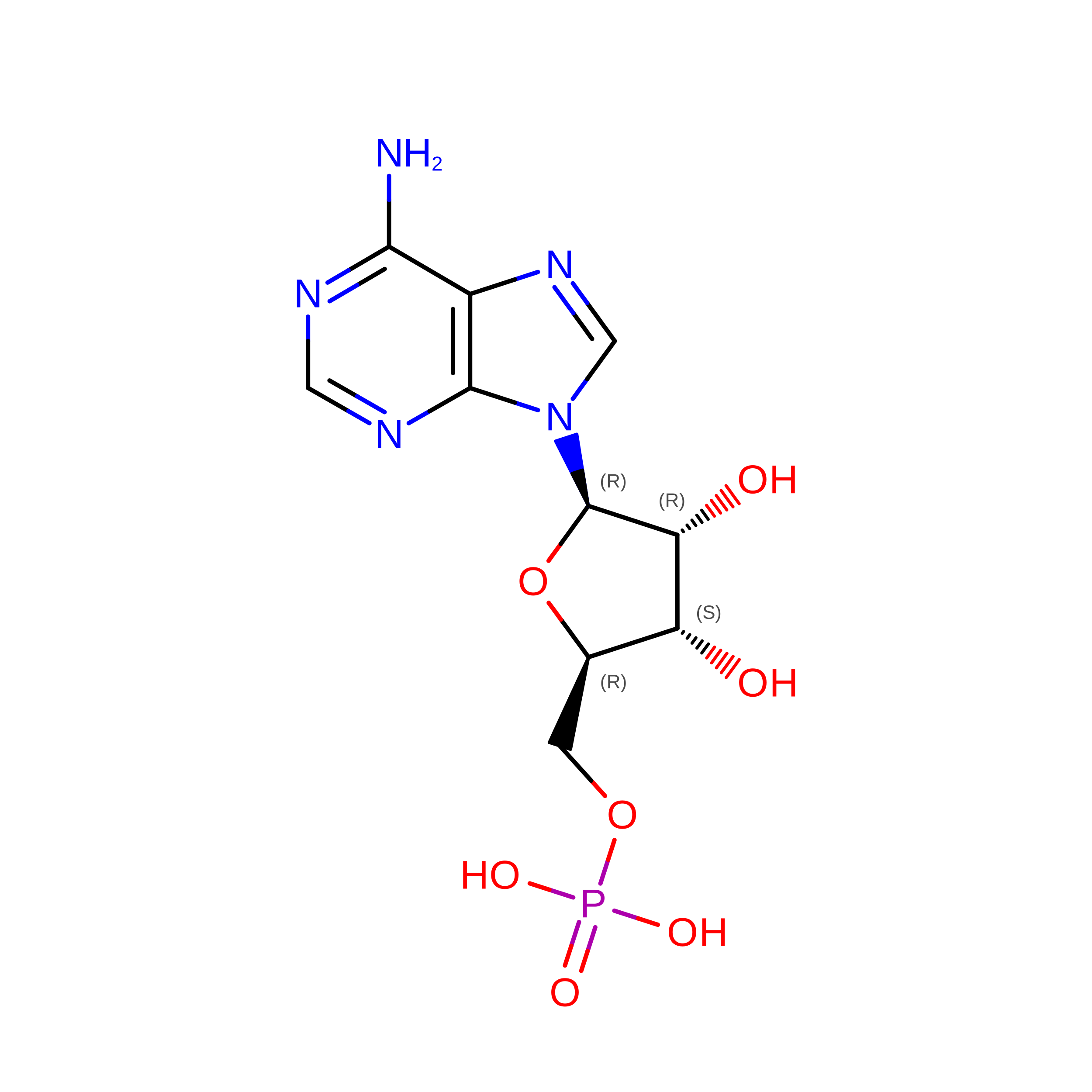
B [auth A]ADENOSINE MONOPHOSPHATE
C10 H14 N5 O7 P
UDMBCSSLTHHNCD-KQYNXXCUSA-N

Experimental Data & Validation

Experimental Data

  • Method: X-RAY DIFFRACTION
  • Resolution: 1.33 Å
  • R-Value Free:  0.207 (Depositor) 
  • R-Value Work:  0.169 (Depositor) 
  • R-Value Observed: 0.171 (Depositor) 
Space Group: C 1 2 1
Unit Cell:
Length ( Å )Angle ( ˚ )
a = 66.132α = 90
b = 43.757β = 118.83
c = 55.654γ = 90
Software Package:
Software NamePurpose
REFMACrefinement
d*TREKdata reduction
d*TREKdata scaling
CCP4phasing

Structure Validation

View Full Validation Report


Ligand Structure Quality Assessment 


Entry History 

Deposition Data

Revision History  (Full details and data files)

  • Version 1.0: 2007-05-08
    Type: Initial release
  • Version 1.1: 2011-05-08
    Changes: Version format compliance
  • Version 1.2: 2011-07-13
    Changes: Version format compliance
  • Version 1.3: 2024-05-08
    Changes: Data collection, Database references, Other