2RUI | pdb_00002rui

Solution Structure of the Bacillus anthracis Sortase A-substrate Complex


Experimental Data Snapshot

  • Method: SOLUTION NMR
  • Conformers Calculated: 200 
  • Conformers Submitted: 20 
  • Selection Criteria: structures with the lowest energy 

wwPDB Validation 3D Report Full Report

Validation slider image for 2RUI

This is version 2.0 of the entry. See complete history

Literature

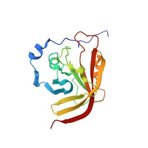
Structure of the Bacillus anthracis Sortase A Enzyme Bound to Its Sorting Signal: A FLEXIBLE AMINO-TERMINAL APPENDAGE MODULATES SUBSTRATE ACCESS.

Chan, A.H.Yi, S.W.Terwilliger, A.L.Maresso, A.W.Jung, M.E.Clubb, R.T.

(2015) J Biological Chem 290: 25461-25474

  • DOI: https://doi.org/10.1074/jbc.M115.670984
  • Primary Citation Related Structures: 
    2RUI

  • PubMed Abstract: 

    The endospore forming bacterium Bacillus anthracis causes lethal anthrax disease in humans and animals. The ability of this pathogen to replicate within macrophages is dependent upon the display of bacterial surface proteins attached to the cell wall by the B. anthracis Sortase A ((Ba)SrtA) enzyme. Previously, we discovered that the class A (Ba)SrtA sortase contains a unique N-terminal appendage that wraps around the body of the protein to contact the active site of the enzyme. To gain insight into its function, we determined the NMR structure of (Ba)SrtA bound to a LPXTG sorting signal analog. The structure, combined with dynamics, kinetics, and whole cell protein display data suggest that the N terminus modulates substrate access to the enzyme. We propose that it may increase the efficiency of protein display by reducing the unproductive hydrolytic cleavage of enzyme-protein covalent intermediates that form during the cell wall anchoring reaction. Notably, a key active site loop (β7/β8 loop) undergoes a disordered to ordered transition upon binding the sorting signal, potentially facilitating recognition of lipid II.


  • Organizational Affiliation
    • From the Department of Chemistry and Biochemistry, UCLA-DOE Institute of Genomics and Proteomics, and the Molecular Biology Institute, University of California, Los Angeles, California 90095 and.

Macromolecule Content 

  • Total Structure Weight: 17.61 kDa 
  • Atom Count: 1,233 
  • Modeled Residue Count: 163 
  • Deposited Residue Count: 163 
  • Unique protein chains: 2

Macromolecules

Find similar proteins by:|  3D Structure
Entity ID: 1
MoleculeChains  Sequence LengthOrganismDetailsImage
LPXTG-site transpeptidase family protein158Bacillus anthracis str. SterneMutation(s): 0 
Gene Names: BAS0654srtA
UniProt
Find proteins for Q81V16 (Bacillus anthracis)
Explore Q81V16 
Go to UniProtKB:  Q81V16
Entity Groups
Sequence Clusters30% Identity50% Identity70% Identity90% Identity95% Identity100% Identity
UniProt GroupQ81V16
Sequence Annotations
Expand
Reference Sequence
Find similar proteins by:  Sequence   |   3D Structure  
Entity ID: 2
MoleculeChains  Sequence LengthOrganismDetailsImage
Boc-LPAT*5synthetic constructMutation(s): 0 

Biologically Interesting Molecules (External Reference) 

1 Unique

Experimental Data & Validation

Experimental Data

  • Method: SOLUTION NMR
  • Conformers Calculated: 200 
  • Conformers Submitted: 20 
  • Selection Criteria: structures with the lowest energy 

Structure Validation

View Full Validation Report


Entry History 

Deposition Data

Revision History  (Full details and data files)

  • Version 1.0: 2015-09-09
    Type: Initial release
  • Version 1.1: 2019-12-25
    Changes: Data collection, Database references, Derived calculations
  • Version 2.0: 2023-11-15
    Changes: Atomic model, Data collection, Database references, Derived calculations